Copyright
©The Author(s) 2021.
World J Clin Cases. Nov 26, 2021; 9(33): 10374-10381
Published online Nov 26, 2021. doi: 10.12998/wjcc.v9.i33.10374
Published online Nov 26, 2021. doi: 10.12998/wjcc.v9.i33.10374
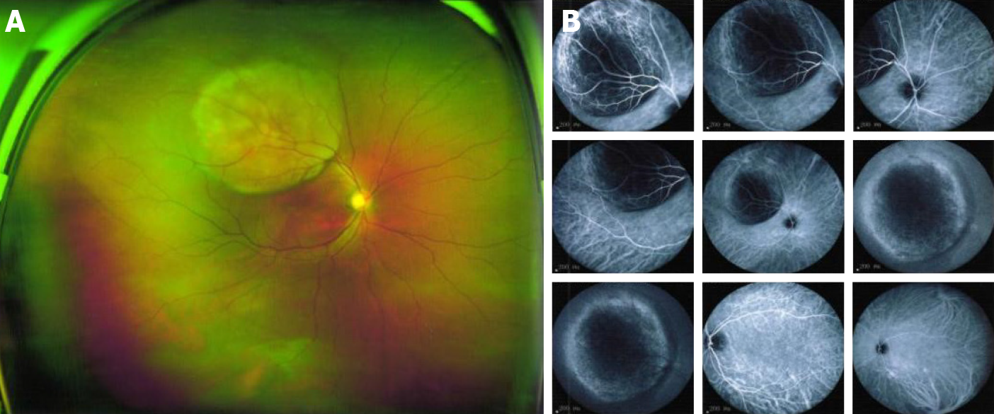
Figure 2 Ophthalmoscopy of the right eye.
A, B: Solid dark gray mass in the posterior segment (choroid) with intense brown pigmentation, occupying posterior third of the vitreous chamber along with mild retinal detachment observed at the peripheral rim of the nodular choroidal mass. The mass size of 11.11 mm × 12.1 mm.
- Citation: Zhang YS, Hu TC, Ye YC, Han JH, Li XJ, Zhang YH, Chen WZ, Chai HY, Pan X, Wang X, Yang YL. Carbon ion radiotherapy for synchronous choroidal melanoma and lung cancer: A case report. World J Clin Cases 2021; 9(33): 10374-10381
- URL: https://www.wjgnet.com/2307-8960/full/v9/i33/10374.htm
- DOI: https://dx.doi.org/10.12998/wjcc.v9.i33.10374


