Copyright
©The Author(s) 2021.
World J Clin Cases. Nov 26, 2021; 9(33): 10345-10354
Published online Nov 26, 2021. doi: 10.12998/wjcc.v9.i33.10345
Published online Nov 26, 2021. doi: 10.12998/wjcc.v9.i33.10345
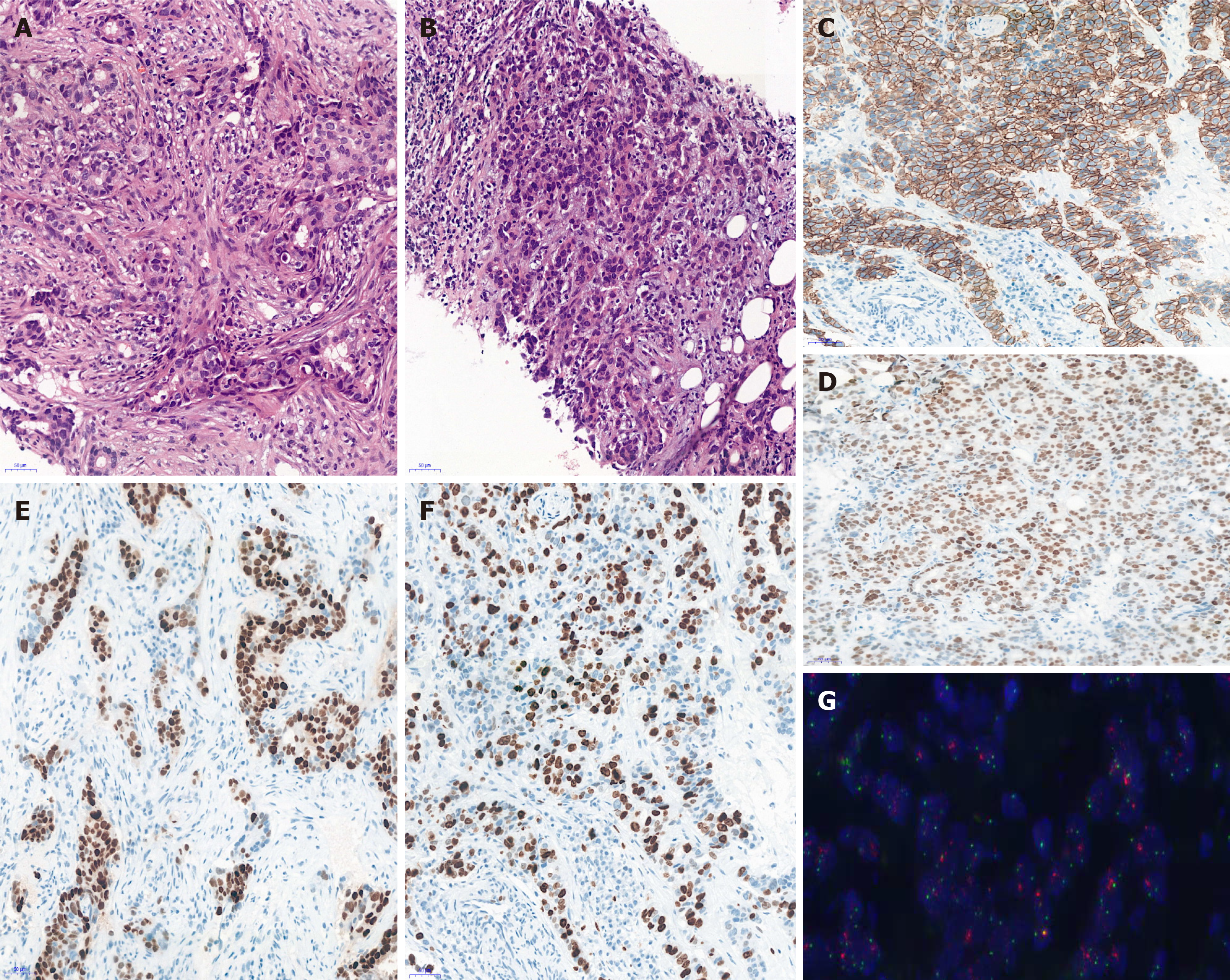
Figure 2 Pathological results of left breast tumor and left axillary lymph node biopsies.
A: Hematoxylin and eosin-stained sections revealed that the tumor cells grew in a solid and patchy infiltrating manner (original magnification: 200 ×); B: Hematoxylin and eosin-stained sections revealed that the left axillary lymph node was metastatic carcinoma, which was consistent with the breast source (original magnification: 200 ×); C: C-erBb2 (2+) was uncertain in neoplastic cells by immunohistochemical analysis (original magnification: 200 ×); D and E: Estrogen receptor and progesterone receptor were negative in neoplastic cells by immunohistochemical analysis (original magnification: 200 ×); F: Ki-67 was expressed in the nuclei of approximately 40% of tumor cells (original magnification: 200 ×); G: HER-2 was amplified by fluorescence in situ hybridization (original magnification: 200 ×).
- Citation: Cai JH, Zheng JH, Lin XQ, Lin WX, Zou J, Chen YK, Li ZY, Chen YX. Individualized treatment of breast cancer with chronic renal failure: A case report and review of literature. World J Clin Cases 2021; 9(33): 10345-10354
- URL: https://www.wjgnet.com/2307-8960/full/v9/i33/10345.htm
- DOI: https://dx.doi.org/10.12998/wjcc.v9.i33.10345


