Copyright
©The Author(s) 2021.
World J Clin Cases. Nov 26, 2021; 9(33): 10308-10314
Published online Nov 26, 2021. doi: 10.12998/wjcc.v9.i33.10308
Published online Nov 26, 2021. doi: 10.12998/wjcc.v9.i33.10308
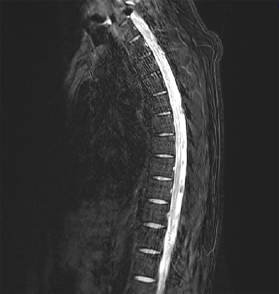
Figure 3 Spine magnetic resonance imaging made on admission.
High signal intensity was apparent within the left spinal cord at level T2-8 on a T2-weighted image.
- Citation: Yun D, Cho SY, Ju W, Seo EH. Transverse myelitis after infection with varicella zoster virus in patient with normal immunity: A case report. World J Clin Cases 2021; 9(33): 10308-10314
- URL: https://www.wjgnet.com/2307-8960/full/v9/i33/10308.htm
- DOI: https://dx.doi.org/10.12998/wjcc.v9.i33.10308


