Copyright
©The Author(s) 2021.
World J Clin Cases. Nov 26, 2021; 9(33): 10286-10292
Published online Nov 26, 2021. doi: 10.12998/wjcc.v9.i33.10286
Published online Nov 26, 2021. doi: 10.12998/wjcc.v9.i33.10286
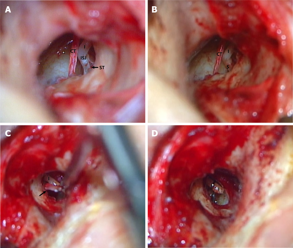
Figure 3 Microscopic examination.
A and B: Microscopic findings of the ossified stapedial tendon and after removal observed during left-sided exploratory tympanotomy; C and D: Microscopic findings of monorod-shaped stapes being removed and after insertion of the piston wire observed during right-sided stapedotomy. CT: Chorda tympani; I: Incus; ISJ: Incudostapedial joint; ST: Stapedial tendon; S: Stapes.
- Citation: Yoo JS, Lee CM, Yang YN, Lee EJ. Complete restoration of congenital conductive hearing loss by staged surgery: A case report. World J Clin Cases 2021; 9(33): 10286-10292
- URL: https://www.wjgnet.com/2307-8960/full/v9/i33/10286.htm
- DOI: https://dx.doi.org/10.12998/wjcc.v9.i33.10286


