Copyright
©The Author(s) 2021.
World J Clin Cases. Nov 26, 2021; 9(33): 10088-10097
Published online Nov 26, 2021. doi: 10.12998/wjcc.v9.i33.10088
Published online Nov 26, 2021. doi: 10.12998/wjcc.v9.i33.10088
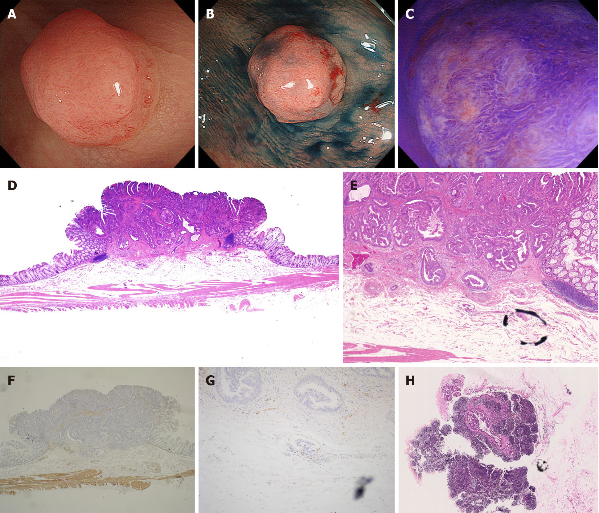
Figure 2 A typical case of small T1 colorectal cancer with lymph node metastasis positivity.
A: An 8-mm-sized lesion of erythematous color located in the sigmoid colon was detected by white light observation; B: Indigo carmine spray observation showed elevation in the center and a depression line at the edge, and was diagnosed as Is + IIc by morphology; C: By magnification observation with crystal violet staining, a non-structured area was identified around severe irregular pits diagnosed as VN type pit pattern; D: Hematoxylin and eosin (H&E) staining showing well to moderately differentiated adenocarcinoma; E: Victoria blue staining. Vascular invasion was positive; F: Desmin staining. Depth of invasion was 3750 μm; G: D2-40 staining. Lymphatic invasion was positive; H: Dissected lymph nodes by H&E staining. Metastasis was positive.
- Citation: Takashina Y, Kudo SE, Ichimasa K, Kouyama Y, Mochizuki K, Akimoto Y, Maeda Y, Mori Y, Misawa M, Ogata N, Kudo T, Hisayuki T, Hayashi T, Wakamura K, Sawada N, Baba T, Ishida F, Yokoyama K, Daita M, Nemoto T, Miyachi H. Clinicopathological features of small T1 colorectal cancers. World J Clin Cases 2021; 9(33): 10088-10097
- URL: https://www.wjgnet.com/2307-8960/full/v9/i33/10088.htm
- DOI: https://dx.doi.org/10.12998/wjcc.v9.i33.10088


