Copyright
©The Author(s) 2021.
World J Clin Cases. Nov 16, 2021; 9(32): 9990-9996
Published online Nov 16, 2021. doi: 10.12998/wjcc.v9.i32.9990
Published online Nov 16, 2021. doi: 10.12998/wjcc.v9.i32.9990
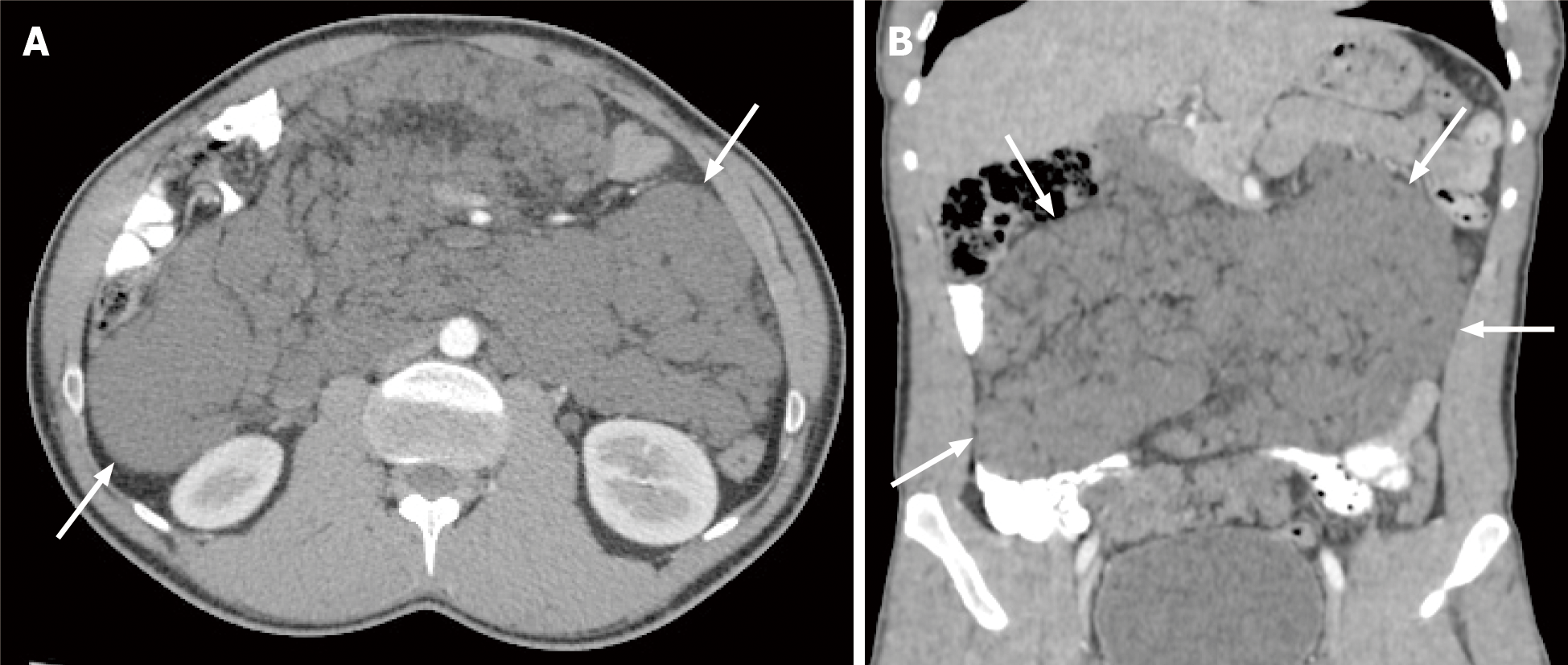
Figure 1 Computed tomography image of confluent peritoneal cystic lesions.
A: Post-contrast axial; B: Coronal computed tomography scan reveals numerous confluent peritoneal cystic lesions (white arrows) of variable sizes extensively involving the small bowel mesentery and responsible for peripheral displacement of the small bowel loops. No retro-peritoneal lesions are seen.
- Citation: Alhasan AS, Daqqaq TS. Extensive abdominal lymphangiomatosis involving the small bowel mesentery: A case report. World J Clin Cases 2021; 9(32): 9990-9996
- URL: https://www.wjgnet.com/2307-8960/full/v9/i32/9990.htm
- DOI: https://dx.doi.org/10.12998/wjcc.v9.i32.9990


