Copyright
©The Author(s) 2021.
World J Clin Cases. Nov 16, 2021; 9(32): 9982-9989
Published online Nov 16, 2021. doi: 10.12998/wjcc.v9.i32.9982
Published online Nov 16, 2021. doi: 10.12998/wjcc.v9.i32.9982
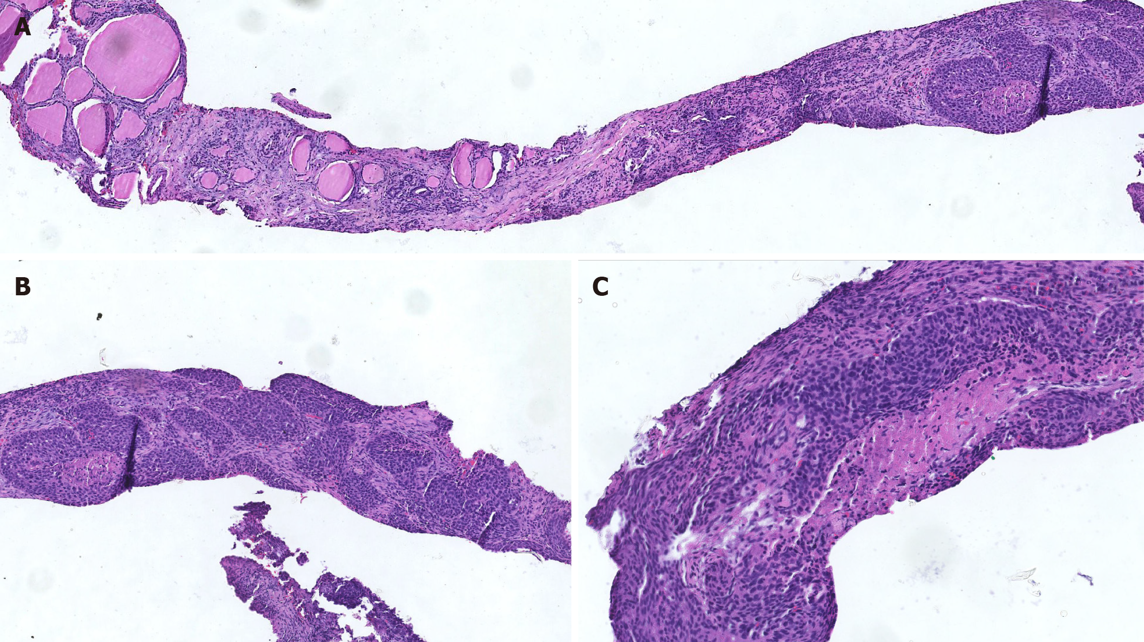
Figure 4 Microscopic features of tissue.
A: Hematoxylin and eosin-stained section of the thyroid mass showing cellular nests of epithelioid cells surrounded by fibrous cells. Toward the end of the tissue, normal thyroid follicles can be observed (×50); B: Tumor cells arranged in nests next to each other in a mosaic pattern with disordered cell polarity (×100); C: Tumor cells with enlarged nuclei with deep staining and rough chromatin, although they are chromatin-rich (×200).
- Citation: Yu JY, Zhang Y, Wang Z. Fine-needle aspiration cytology of an intrathyroidal nodule diagnosed as squamous cell carcinoma: A case report. World J Clin Cases 2021; 9(32): 9982-9989
- URL: https://www.wjgnet.com/2307-8960/full/v9/i32/9982.htm
- DOI: https://dx.doi.org/10.12998/wjcc.v9.i32.9982


