Copyright
©The Author(s) 2021.
World J Clin Cases. Nov 16, 2021; 9(32): 9982-9989
Published online Nov 16, 2021. doi: 10.12998/wjcc.v9.i32.9982
Published online Nov 16, 2021. doi: 10.12998/wjcc.v9.i32.9982
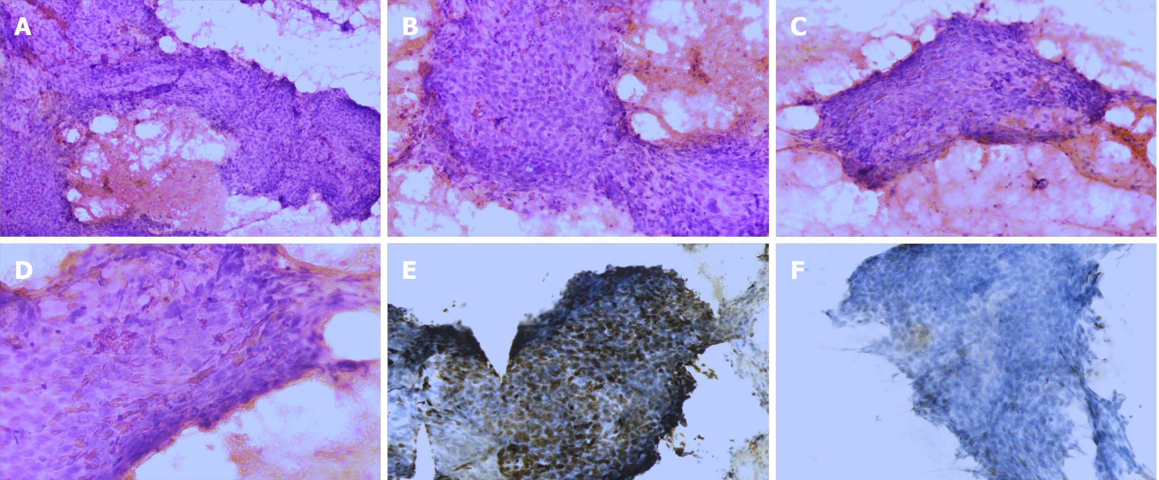
Figure 2 Microscopic features of cytology.
A: Fine-needle aspiration of the thyroid mass showing a large number of neoplastic cells and red blood cells in the background. The neoplastic cells show poor adhesion and are oval or spindle-shaped. These cells appear to be two to five times the size of lymphocytes. They are arranged densely and are disordered (×100); B-D: Image under a high-power objective lens showing neoplastic cells with coarse nuclear chromatin with occasional prominent nucleoli and the cells are rich in cytoplasm. Mitosis can be observed occasionally (×200); E: Tumor cells showing positivity for p63 (×200). F: Tumor cells showing negativity for TTF-1 (×200).
- Citation: Yu JY, Zhang Y, Wang Z. Fine-needle aspiration cytology of an intrathyroidal nodule diagnosed as squamous cell carcinoma: A case report. World J Clin Cases 2021; 9(32): 9982-9989
- URL: https://www.wjgnet.com/2307-8960/full/v9/i32/9982.htm
- DOI: https://dx.doi.org/10.12998/wjcc.v9.i32.9982


