Copyright
©The Author(s) 2021.
World J Clin Cases. Nov 16, 2021; 9(32): 9948-9953
Published online Nov 16, 2021. doi: 10.12998/wjcc.v9.i32.9948
Published online Nov 16, 2021. doi: 10.12998/wjcc.v9.i32.9948
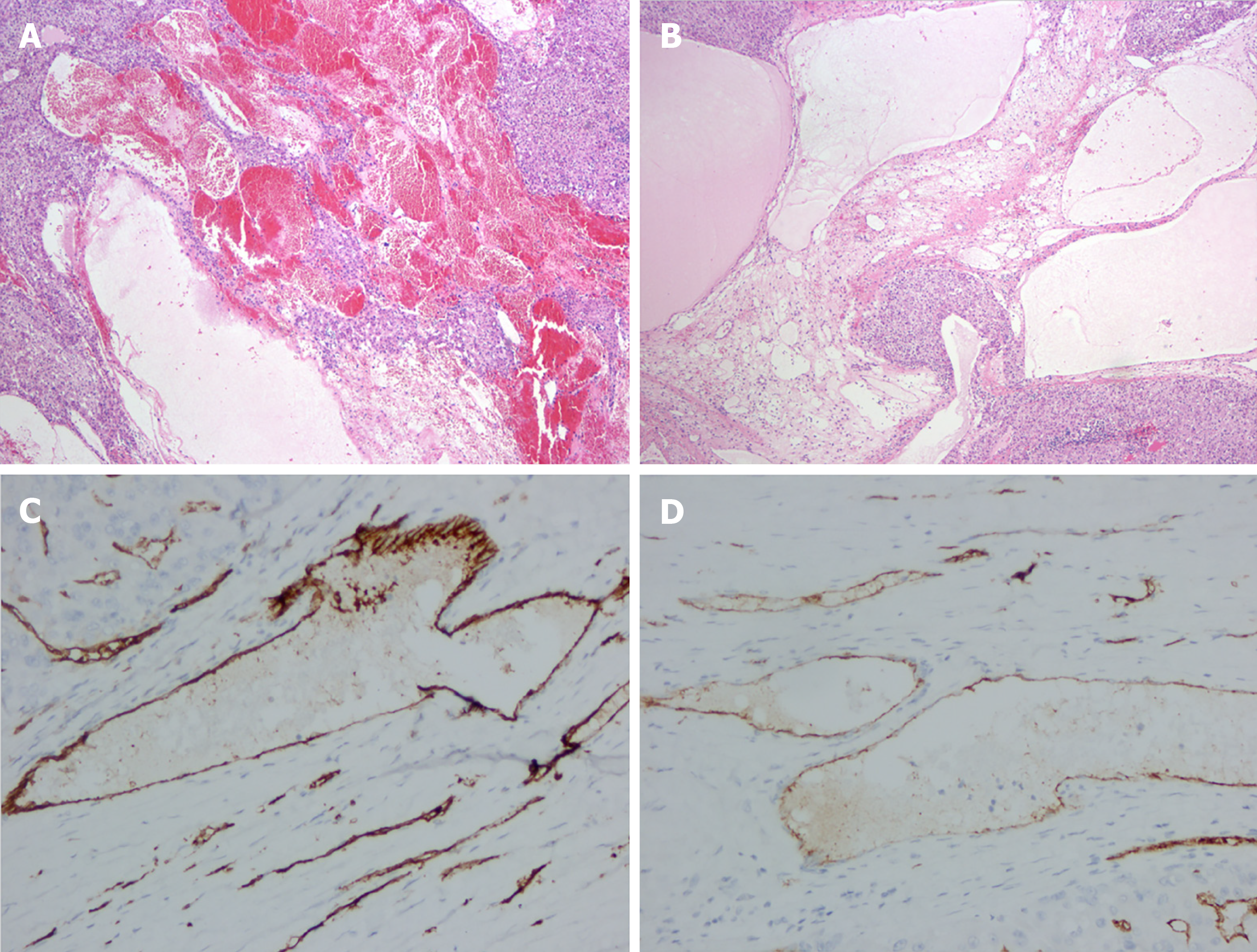
Figure 2 Pathological results of hepatic hemolymphangioma.
A, B: Microscopically, the tumor was mainly composed of lymphatic and blood vessels (40×); C, D: Immunohistochemical staining showed that CD34 and CD31 were positive (400×).
- Citation: Wang M, Liu HF, Zhang YZZ, Zou ZQ, Wu ZQ. Hemolymphangioma with multiple hemangiomas in liver of elderly woman with history of gynecological malignancy: A case report. World J Clin Cases 2021; 9(32): 9948-9953
- URL: https://www.wjgnet.com/2307-8960/full/v9/i32/9948.htm
- DOI: https://dx.doi.org/10.12998/wjcc.v9.i32.9948


