Copyright
©The Author(s) 2021.
World J Clin Cases. Nov 16, 2021; 9(32): 9926-9934
Published online Nov 16, 2021. doi: 10.12998/wjcc.v9.i32.9926
Published online Nov 16, 2021. doi: 10.12998/wjcc.v9.i32.9926
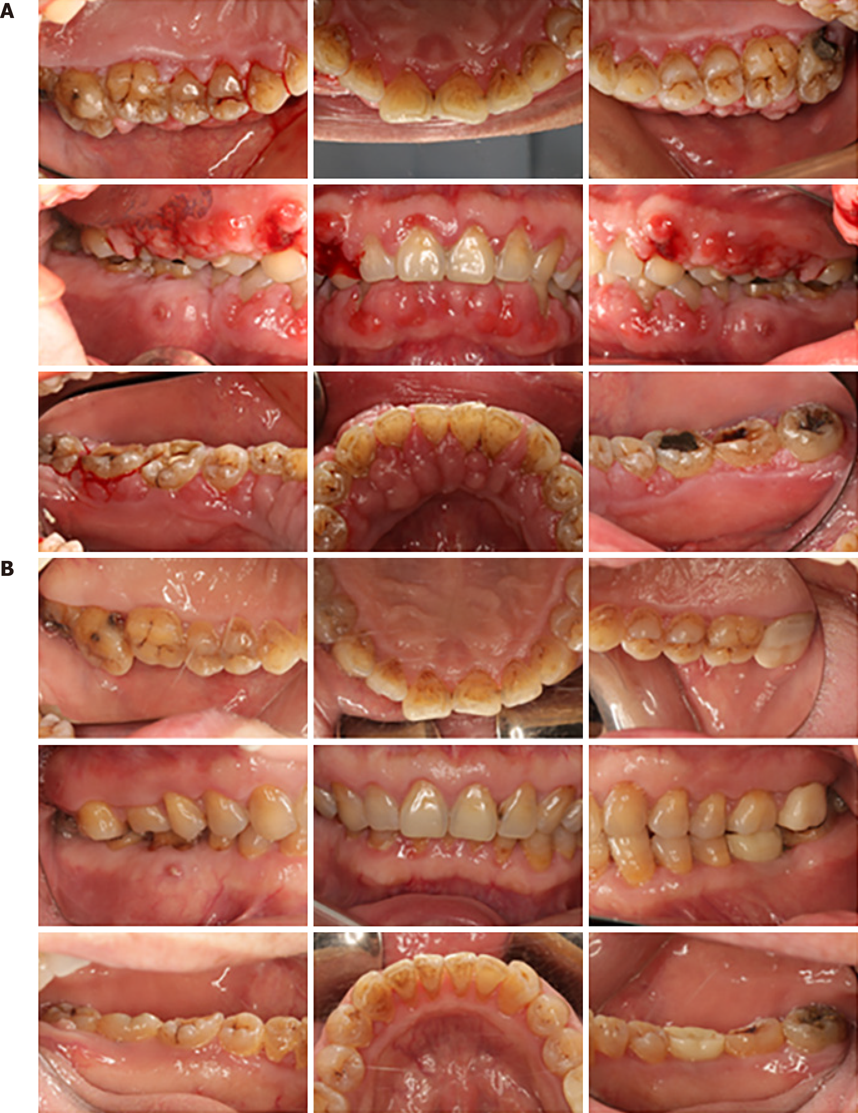
Figure 1 Imaging examination photos.
A: Initial clinical report on Patient 1 showing severe inflammatory and fibrotic GO with a spontaneous bleeding tendency. Fistula over tooth #46, the lower right first molar. A large silver–mercury filling in the crown of tooth #36. Tooth #27 had a large area of crown caries; B: Clinical re-evaluation after 2.5 years. The gingivae were less edematous and glazed. The fistula did not heal after root canal treatment in tooth #46. Later on, the patient requested removal of the affected tooth. The crown was repaired after root canal treatment of tooth #27. Tooth #36 was extracted due to fracture, and a dental implant was given.
- Citation: Fang L, Tan BC. Clinical presentation and management of drug-induced gingival overgrowth: A case series. World J Clin Cases 2021; 9(32): 9926-9934
- URL: https://www.wjgnet.com/2307-8960/full/v9/i32/9926.htm
- DOI: https://dx.doi.org/10.12998/wjcc.v9.i32.9926


