Copyright
©The Author(s) 2021.
World J Clin Cases. Nov 16, 2021; 9(32): 9889-9895
Published online Nov 16, 2021. doi: 10.12998/wjcc.v9.i32.9889
Published online Nov 16, 2021. doi: 10.12998/wjcc.v9.i32.9889
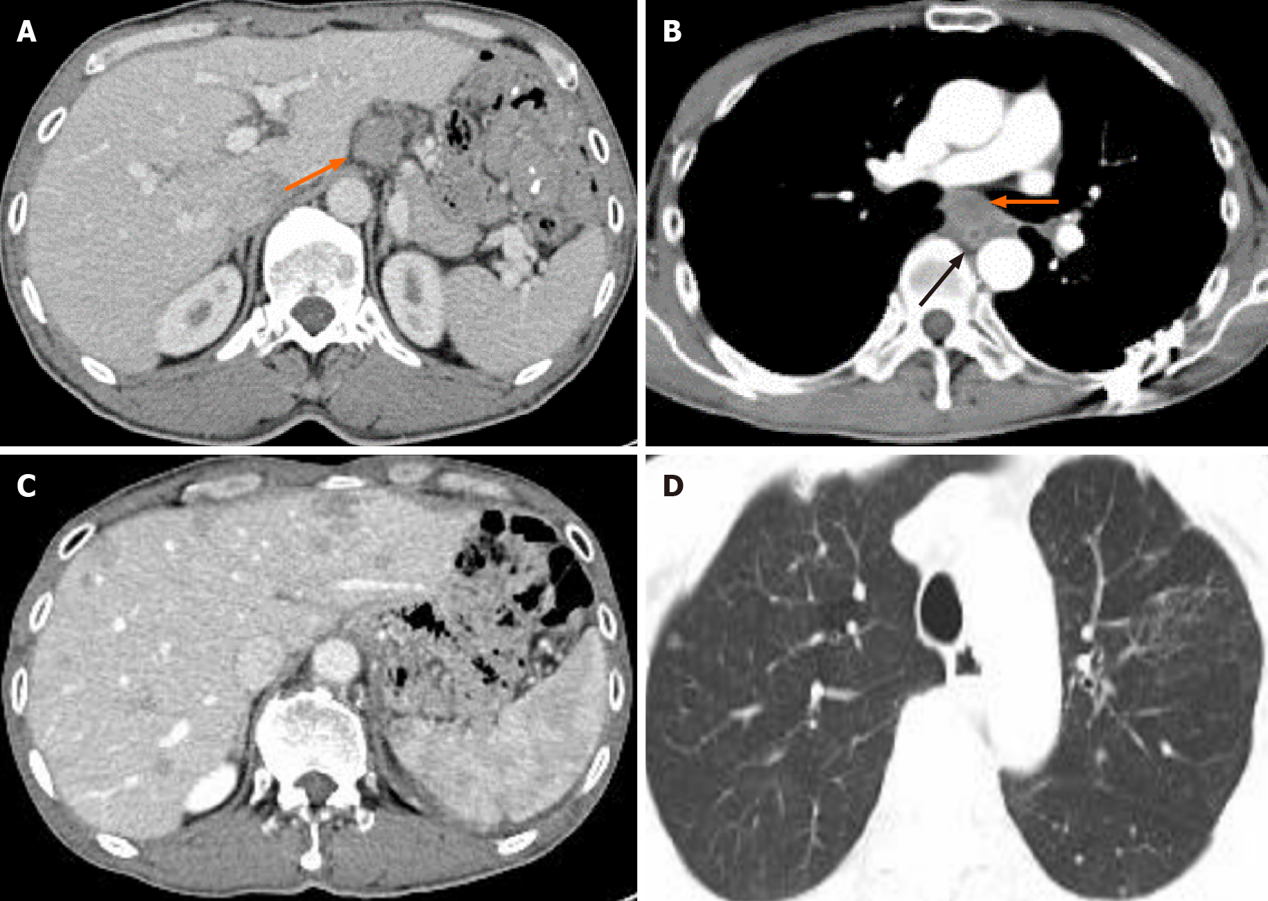
Figure 4 Computed tomography re-examination.
A: Computed tomography (CT) re-examination showed that the hepatogastric ligamentous lymph node enlarged 21 mo after surgery (orange arrow); B: CT re-examination showed that the local thickening of the esophageal anastomotic site (black arrow) and enlarged subcarinal lymph nodes (orange arrow); C: CT re-examination showed multiple nodules in the liver; D: CT re-examination showed multiple nodules scattered in both lungs emerging 28 mo after surgery.
- Citation: Li Y, Ye LS, Hu B. Synchronous multiple primary malignancies of the esophagus, stomach, and jejunum: A case report. World J Clin Cases 2021; 9(32): 9889-9895
- URL: https://www.wjgnet.com/2307-8960/full/v9/i32/9889.htm
- DOI: https://dx.doi.org/10.12998/wjcc.v9.i32.9889


