Copyright
©The Author(s) 2021.
World J Clin Cases. Nov 16, 2021; 9(32): 10033-10039
Published online Nov 16, 2021. doi: 10.12998/wjcc.v9.i32.10033
Published online Nov 16, 2021. doi: 10.12998/wjcc.v9.i32.10033
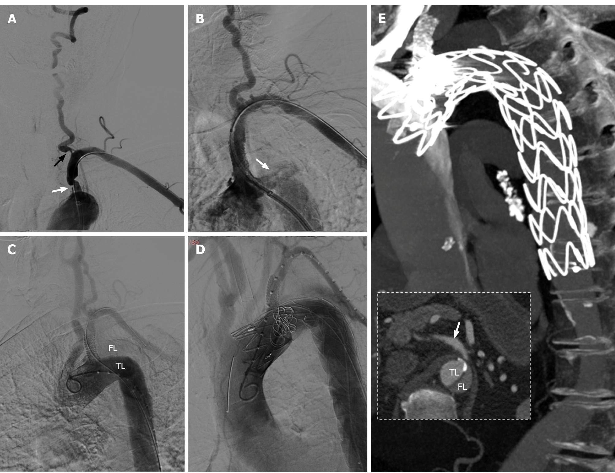
Figure 3 The procedures of subclavian artery stenting for stenosis and thoracic endovascular aortic repair for aortic dissection.
A: Severe stenosis of the proximal left subclavian artery (LSA) (white arrow) and the ostium of ipsilateral vertebral artery (black arrow) were presented in digital subtraction angiography; B: Contrast entering the aortic wall (white arrow) after a balloon expandable stent implanted into LSA; C: The true lumen and false lumen (FL) could be clearly displayed in angiogram of aortic dissection, and the origin of dissection was identified at the proximal edge of stent; D: Chimney technique was used in thoracic endovascular aortic repair; E: Two-week computed tomography angiography (CTA) follow-up, there was no significant contrast enter into the FL, and the distal end of aortic dissection was at least below the bifurcation point of celiac trunk (white arrow, CTA failed to scan the whole dissection).
- Citation: Zhang Y, Wang JW, Jin G, Liang B, Li X, Yang YT, Zhan QL. Focal intramural hematoma as a potential pitfall for iatrogenic aortic dissection during subclavian artery stenting: A case report. World J Clin Cases 2021; 9(32): 10033-10039
- URL: https://www.wjgnet.com/2307-8960/full/v9/i32/10033.htm
- DOI: https://dx.doi.org/10.12998/wjcc.v9.i32.10033


