Copyright
©The Author(s) 2021.
World J Clin Cases. Nov 16, 2021; 9(32): 10033-10039
Published online Nov 16, 2021. doi: 10.12998/wjcc.v9.i32.10033
Published online Nov 16, 2021. doi: 10.12998/wjcc.v9.i32.10033
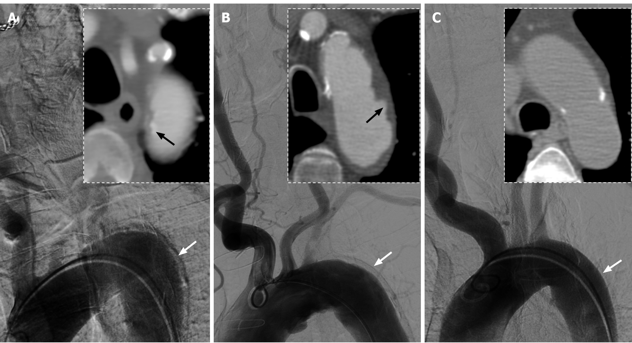
Figure 2 Comparison of aortic arch images between three male patients with anteroposterior views on digital subtraction angiography and axial views on computed tomography angiography.
A: Images of this present case showed a significant artifact on lateral wall of descending aorta (white arrow) and displacement of aortic intimal calcification (black arrow); B: Images of the second patient, aged 79-years-old, showed a similar artifact (white arrow) and penetrating atherosclerotic ulcers (black arrow); C: Images of the third patient, aged 64-years-old, showed mild atherosclerosis on the aortic wall without obvious artifact (white arrow).
- Citation: Zhang Y, Wang JW, Jin G, Liang B, Li X, Yang YT, Zhan QL. Focal intramural hematoma as a potential pitfall for iatrogenic aortic dissection during subclavian artery stenting: A case report. World J Clin Cases 2021; 9(32): 10033-10039
- URL: https://www.wjgnet.com/2307-8960/full/v9/i32/10033.htm
- DOI: https://dx.doi.org/10.12998/wjcc.v9.i32.10033


