Copyright
©The Author(s) 2021.
World J Clin Cases. Nov 6, 2021; 9(31): 9670-9679
Published online Nov 6, 2021. doi: 10.12998/wjcc.v9.i31.9670
Published online Nov 6, 2021. doi: 10.12998/wjcc.v9.i31.9670
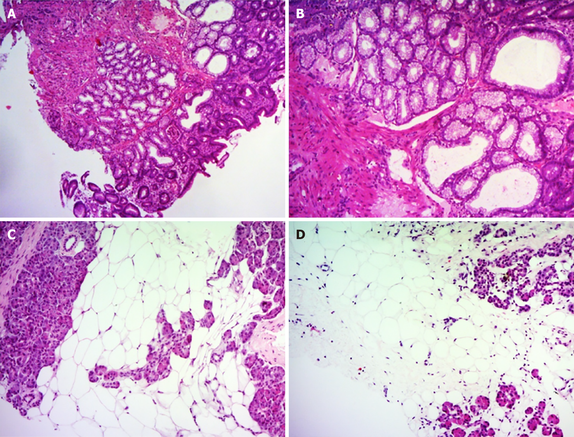
Figure 4 Histological findings of duodenal and pancreatic biopsy specimens.
A and B: The duodenal biopsy showed lobulated proliferation of submucosal Brunner’ glands comprising benign-looking acini lined by mucous cells with basal nuclei without atypia, which was consistent with Brunner’s gland hyperplasia. C and D: Percutaneous pancreatic biopsies revealed adipose tissue replacing the pancreatic parenchyma. Some pancreatic acini were identified with a scattered distribution. Hematoxylin and eosin stain (A–D). Original magnification: (A, C, D) × 40, (B) × 100.
- Citation: Nguyen LC, Vu KT, Vo TTT, Trinh CH, Do TD, Pham NTV, Pham TV, Nguyen TT, Nguyen HC, Byeon JS. Brunner’s gland hyperplasia associated with lipomatous pseudohypertrophy of the pancreas presenting with gastrointestinal bleeding: A case report. World J Clin Cases 2021; 9(31): 9670-9679
- URL: https://www.wjgnet.com/2307-8960/full/v9/i31/9670.htm
- DOI: https://dx.doi.org/10.12998/wjcc.v9.i31.9670


