Copyright
©The Author(s) 2021.
World J Clin Cases. Nov 6, 2021; 9(31): 9652-9661
Published online Nov 6, 2021. doi: 10.12998/wjcc.v9.i31.9652
Published online Nov 6, 2021. doi: 10.12998/wjcc.v9.i31.9652
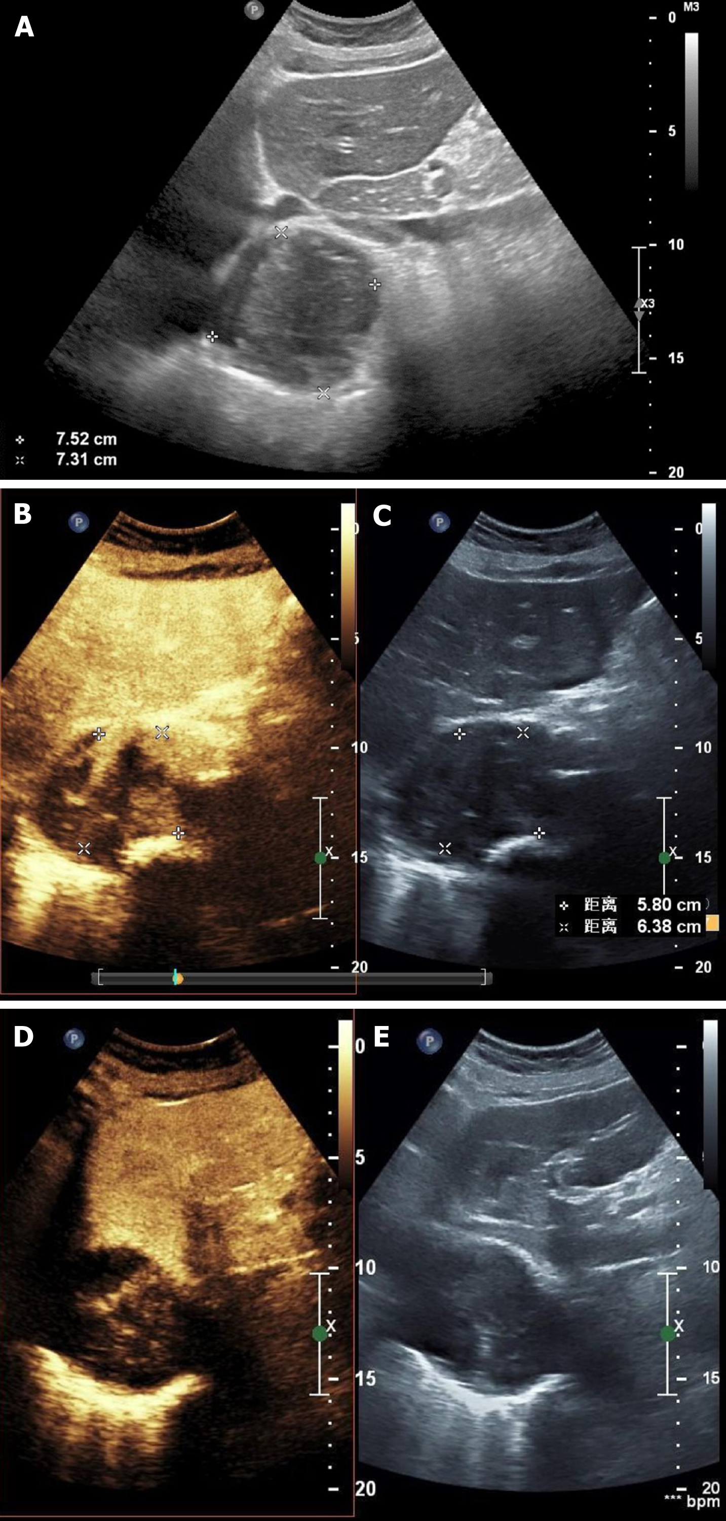
Figure 2 Ultrasound and contrast-enhanced ultrasonography images.
A: Two-dimensional ultrasound image. The marked circular area is the mass; B: Synchronous contrast-enhanced ultrasonography (CEUS) pictures showing the harmonic mode; C: Synchronous CEUS pictures showing the basal mode; D: Synchronous CEUS pictures at different times and levels compared to B and C in the harmonic mode, in which the contrast signal was highlighted; E: Synchronous CEUS pictures at different times and levels compared to B and C in the basal mode, in which only the tissue signal was displayed similar to a normal ultrasound and the contrast agent signal was not highlighted. Solids have stronger properties reflecting ultrasound than liquid material, which present as a stronger signal and show more brightness in the basal mode picture. In the harmonic mode, the higher the content of contrast agent in the tissue, the stronger the signal and the higher the brightness in the image. As seen in panel A, the echo of the mass around the artery was uneven and was comprised of liquid anechoic and solid hyperechoic areas. By comparing panels B and C with panels D and E, it can be observed that the contrast medium predominantly diffusely filled the hyperechoic areas, rather than the hypoechoic areas. There was no enhancement signal of the contrast medium in the liquid anechoic area but a rich signal of contrast medium in the solid hyperechoic area was present, suggesting that the hyperechoic regions in this study were solid tissues rich in blood supply and capillaries. The lesion was similar to a malignant tumor with liquefaction and necrosis.
- Citation: Xie XJ, Jiang TA, Zhao QY. Diagnostic value of contrast-enhanced ultrasonography in mediastinal leiomyosarcoma mimicking aortic hematoma: A case report and review of literature . World J Clin Cases 2021; 9(31): 9652-9661
- URL: https://www.wjgnet.com/2307-8960/full/v9/i31/9652.htm
- DOI: https://dx.doi.org/10.12998/wjcc.v9.i31.9652


