Copyright
©The Author(s) 2021.
World J Clin Cases. Nov 6, 2021; 9(31): 9598-9606
Published online Nov 6, 2021. doi: 10.12998/wjcc.v9.i31.9598
Published online Nov 6, 2021. doi: 10.12998/wjcc.v9.i31.9598
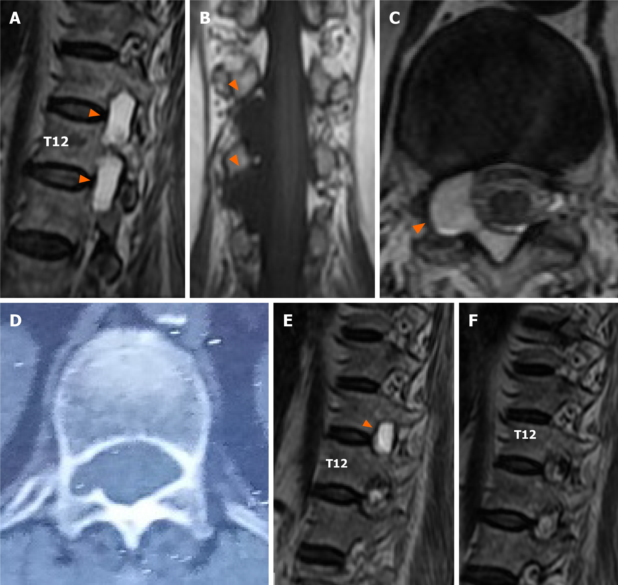
Figure 1 Magnetic resonance imaging and computed tomography scan.
A-C: Preoperative magnetic resonance imaging (MRI) showed cystic lesions (white arrows) at T11-L1 level, T1WI demonstrated a low-intensity signal, and T2WI demonstrated a high-intensity signal. The cyst caused spinal cord compression, moving to the left. Coronal MRI showed that the cystic lesions were in the nerve root (white arrows) axilla at the level of T11-L1; D: Preoperative computed tomography scan revealed bone erosion (white arrows), foraminal enlargement, and enlargement of the spinal canal; E: MRI after the first surgery showed the cyst at T11-T12 (white arrows); F: 2-year follow-up MRI showed no recurrence.
- Citation: Yun ZH, Zhang J, Wu JP, Yu T, Liu QY. Transforaminal endoscopic excision of bi-segmental non-communicating spinal extradural arachnoid cysts: A case report and literature review. World J Clin Cases 2021; 9(31): 9598-9606
- URL: https://www.wjgnet.com/2307-8960/full/v9/i31/9598.htm
- DOI: https://dx.doi.org/10.12998/wjcc.v9.i31.9598


