Copyright
©The Author(s) 2021.
World J Clin Cases. Nov 6, 2021; 9(31): 9584-9591
Published online Nov 6, 2021. doi: 10.12998/wjcc.v9.i31.9584
Published online Nov 6, 2021. doi: 10.12998/wjcc.v9.i31.9584
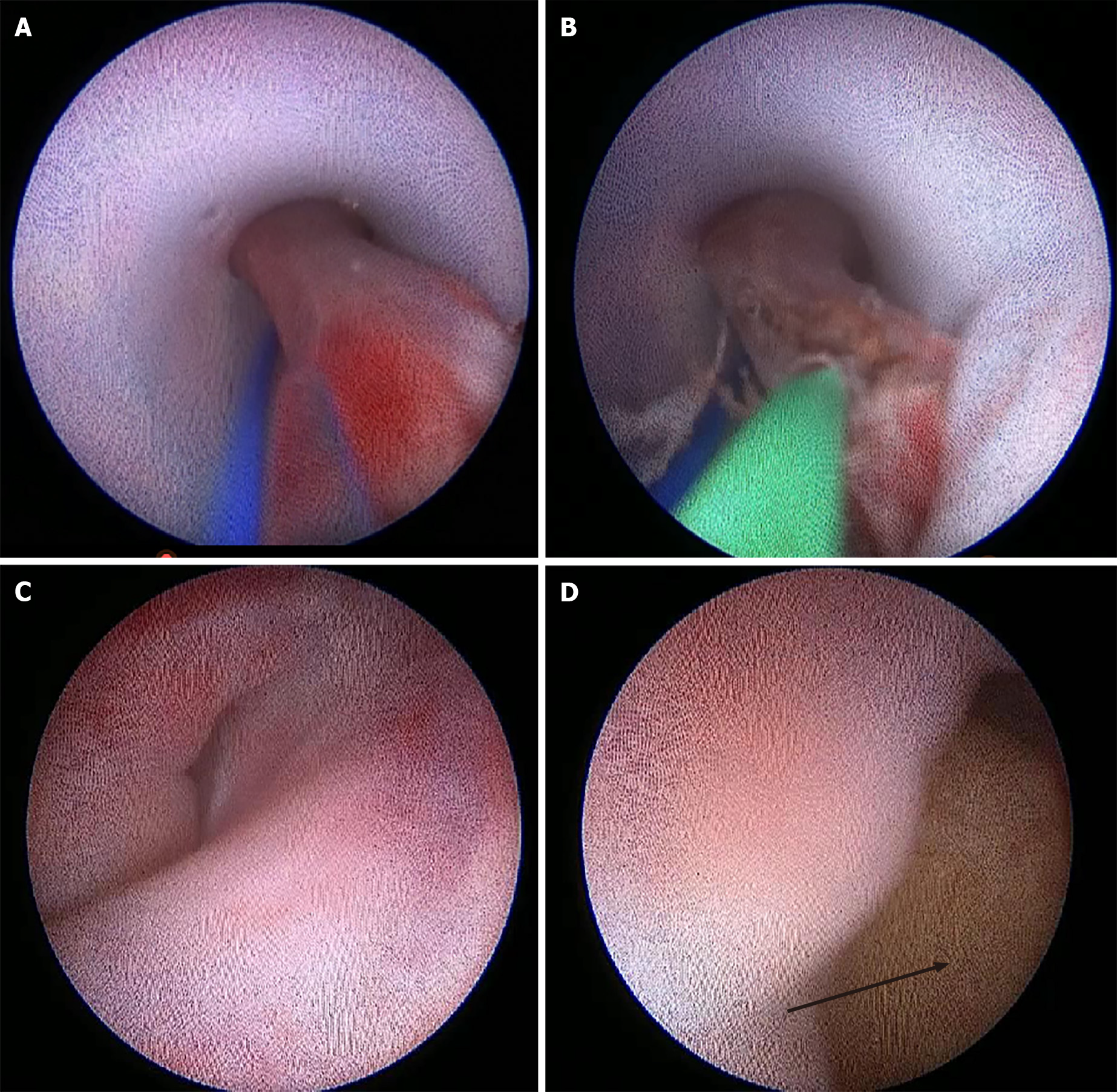
Figure 3 The greater omentum is incarcerated in the peritoneal drain and was cut using a holmium laser under ureteroscopy.
A: The greater omentum protrudes into the drain through the side hole and is incarcerated; B: The greater omentum is transected by the holmium laser fiber; C and D: Show the conditions of the sinus tract and abdominal cavity examined by ureteroscopy after drain removal through the drainage sinus tract, and no bleeding is observed (arrow indicates the greater omentum in the abdominal cavity).
- Citation: Liu HM, Luo GH, Yang XF, Chu ZG, Ye T, Su ZY, Kai L, Yang XS, Wang Z. Ureteroscopic holmium laser to transect the greater omentum to remove an abdominal drain: Four case reports . World J Clin Cases 2021; 9(31): 9584-9591
- URL: https://www.wjgnet.com/2307-8960/full/v9/i31/9584.htm
- DOI: https://dx.doi.org/10.12998/wjcc.v9.i31.9584


