Copyright
©The Author(s) 2021.
World J Clin Cases. Nov 6, 2021; 9(31): 9564-9570
Published online Nov 6, 2021. doi: 10.12998/wjcc.v9.i31.9564
Published online Nov 6, 2021. doi: 10.12998/wjcc.v9.i31.9564
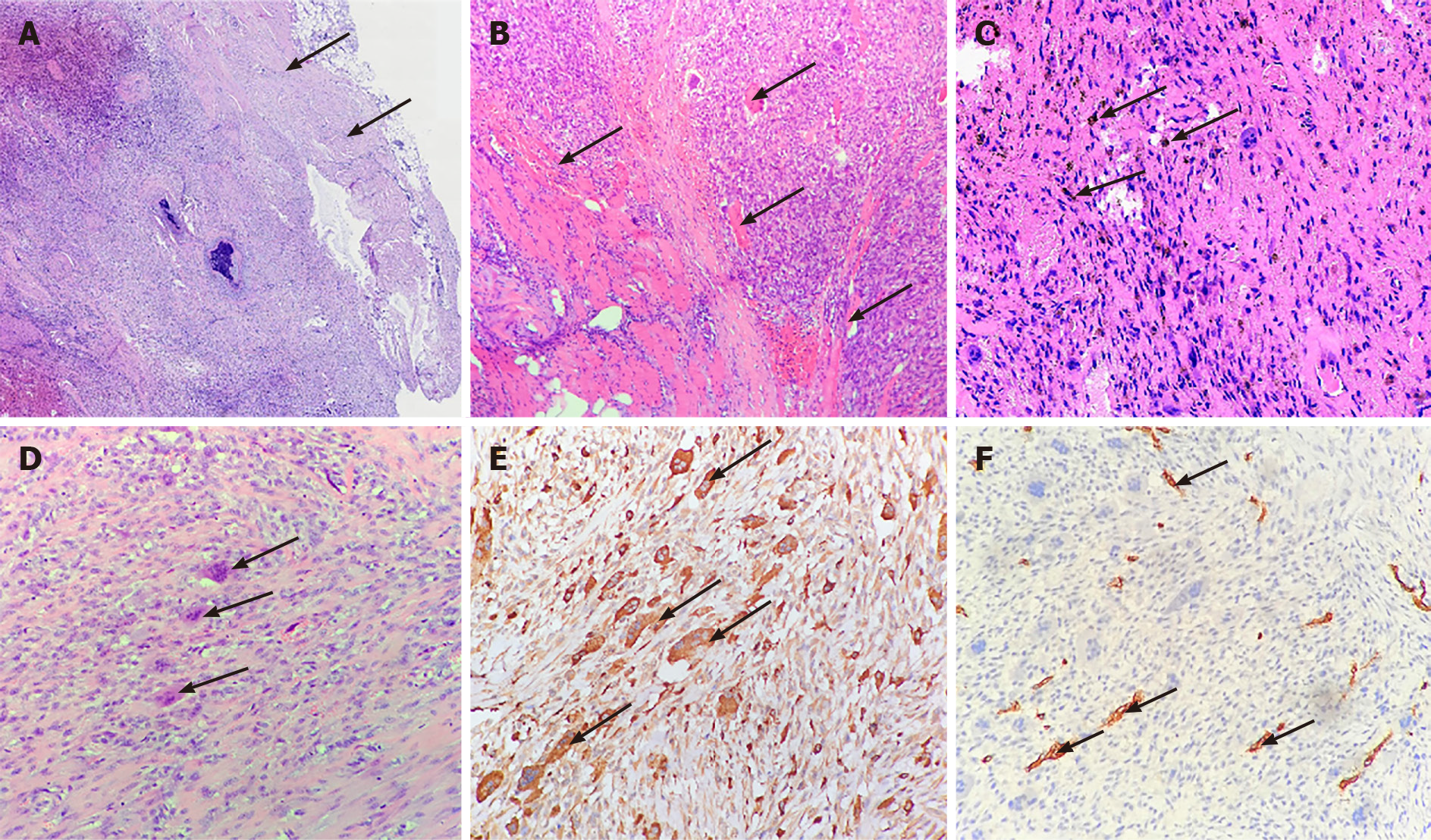
Figure 3 Pathological features of the tumor.
A: The relatively clear boundary (arrows) of the tumor; B: The tumor had invaded neighboring muscles (arrows) on one side; C: Hemosiderin-containing cells (arrows) indicated the tumor with interstitial hemorrhage; D: The tumor had many multinucleated giant cells (arrows) surrounded by spindle cells; E: Multinuclear cells showed strongly positive CD68 (arrows); F: The tumor cells showed negative CD34 but were reactive to vascular endothelial cells (arrows) in the stroma.
- Citation: Kang JY, Zhang K, Liu AL, Wang HL, Zhang LN, Liu WV. Characteristics of primary giant cell tumor in soft tissue on magnetic resonance imaging: A case report. World J Clin Cases 2021; 9(31): 9564-9570
- URL: https://www.wjgnet.com/2307-8960/full/v9/i31/9564.htm
- DOI: https://dx.doi.org/10.12998/wjcc.v9.i31.9564


