Copyright
©The Author(s) 2021.
World J Clin Cases. Nov 6, 2021; 9(31): 9564-9570
Published online Nov 6, 2021. doi: 10.12998/wjcc.v9.i31.9564
Published online Nov 6, 2021. doi: 10.12998/wjcc.v9.i31.9564
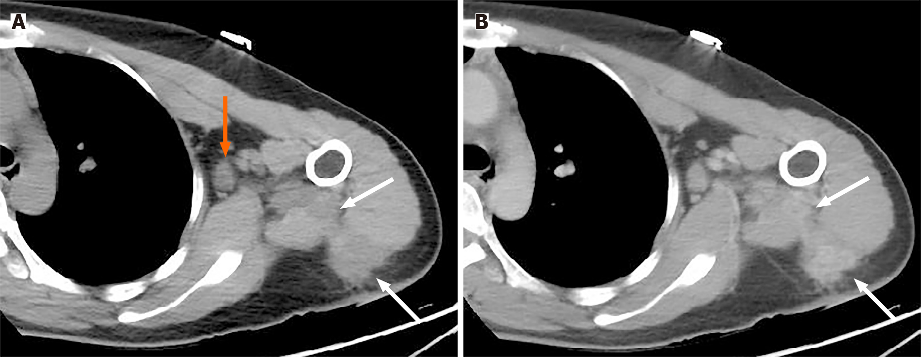
Figure 1 A 36-year-old woman was admitted to our hospital with swelling, skin redness and pain in the upper limb that persisted for 6 mo without a prior history of trauma.
A: Plain computed tomography (CT) imaging shows an irregular hypodense mass in the superficial deltoid muscle extending to the intermuscular space (white arrow) with axillary lymphadenopathy (orange arrow); B: Contrast CT (venous phase) reveals a slightly persistent inhomogeneous enhancement of the mass with a blurred margin (white arrow).
- Citation: Kang JY, Zhang K, Liu AL, Wang HL, Zhang LN, Liu WV. Characteristics of primary giant cell tumor in soft tissue on magnetic resonance imaging: A case report. World J Clin Cases 2021; 9(31): 9564-9570
- URL: https://www.wjgnet.com/2307-8960/full/v9/i31/9564.htm
- DOI: https://dx.doi.org/10.12998/wjcc.v9.i31.9564


