Copyright
©The Author(s) 2021.
World J Clin Cases. Oct 26, 2021; 9(30): 9244-9254
Published online Oct 26, 2021. doi: 10.12998/wjcc.v9.i30.9244
Published online Oct 26, 2021. doi: 10.12998/wjcc.v9.i30.9244
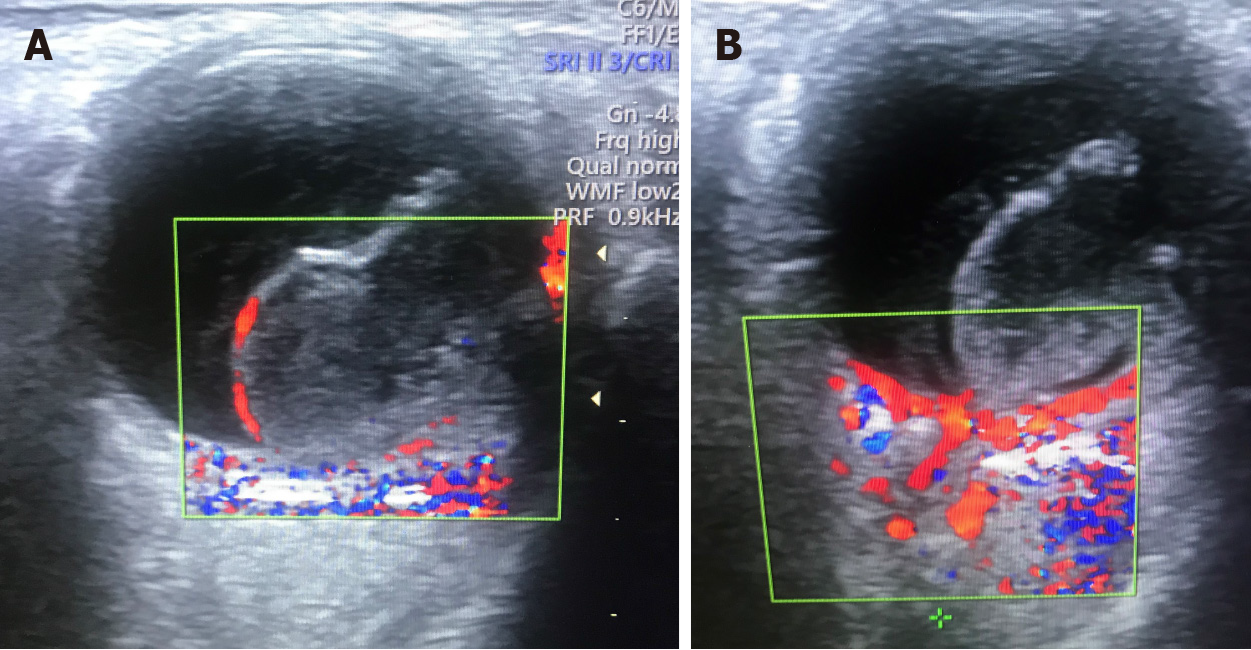
Figure 6 Color Doppler ultrasound examination of the left eye.
A: Blood flow signals were seen in the entire periphery of the occupying lesion; B: There were no obvious blood flow signals in the solid portion in the middle of the lesion.
- Citation: Li Z, Gao W, Tian YM, Xiao Y. Choroidal metastatic mucinous abscess caused by Pseudomonas aeruginosa: A case report. World J Clin Cases 2021; 9(30): 9244-9254
- URL: https://www.wjgnet.com/2307-8960/full/v9/i30/9244.htm
- DOI: https://dx.doi.org/10.12998/wjcc.v9.i30.9244


