Copyright
©The Author(s) 2021.
World J Clin Cases. Oct 26, 2021; 9(30): 9182-9191
Published online Oct 26, 2021. doi: 10.12998/wjcc.v9.i30.9182
Published online Oct 26, 2021. doi: 10.12998/wjcc.v9.i30.9182
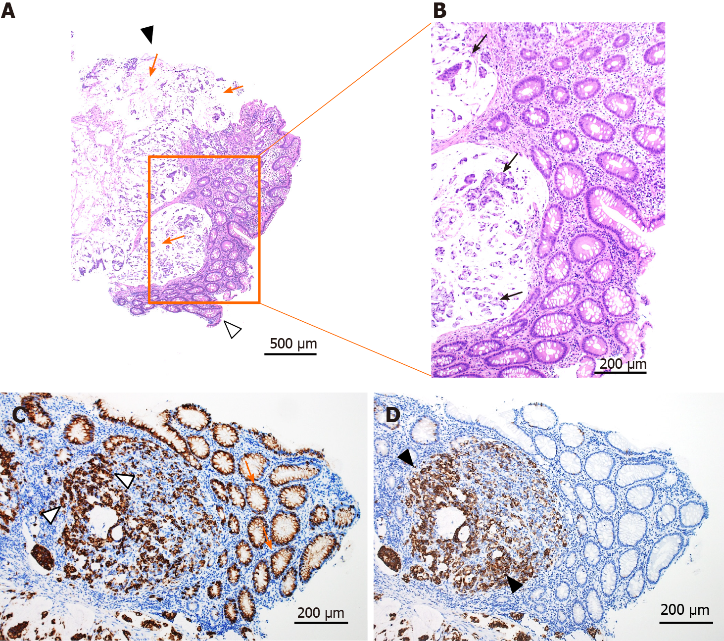
Figure 3 Histological findings of the tumor.
A and B: Hematoxylin and eosin staining. Mucus in the tumor (orange arrows) and adenocarcinoma cells in the mucus (black arrows). Black arrowhead shows the surface of the tumor and white arrowhead shows the bottom of the tumor; C: Mucin 2, oligomeric mucus gel-forming staining (orange arrows and white arrowheads represent positively stained mucosal cells and adenocarcinoma cells); D: Mucin 5AC staining (black arrowheads represent positively stained adenocarcinoma cells).
- Citation: Koseki Y, Kamimura K, Tanaka Y, Ohkoshi-Yamada M, Zhou Q, Matsumoto Y, Mizusawa T, Sato H, Sakamaki A, Umezu H, Yokoyama J, Terai S. Rapid progression of colonic mucinous adenocarcinoma with immunosuppressive condition: A case report and review of literature. World J Clin Cases 2021; 9(30): 9182-9191
- URL: https://www.wjgnet.com/2307-8960/full/v9/i30/9182.htm
- DOI: https://dx.doi.org/10.12998/wjcc.v9.i30.9182


