Copyright
©The Author(s) 2021.
World J Clin Cases. Oct 26, 2021; 9(30): 9182-9191
Published online Oct 26, 2021. doi: 10.12998/wjcc.v9.i30.9182
Published online Oct 26, 2021. doi: 10.12998/wjcc.v9.i30.9182
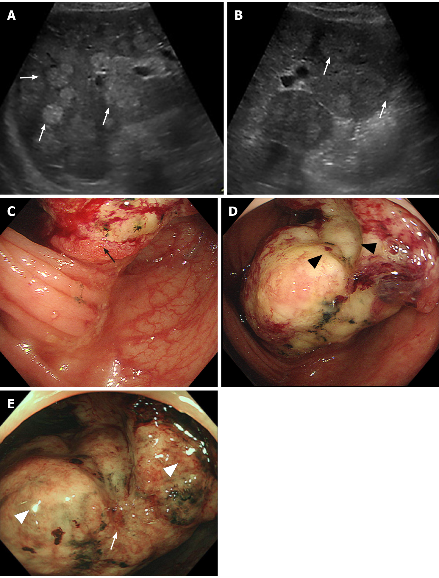
Figure 2 Ultrasonographic and endoscopic images of the tumor.
A and B: Abdominal ultrasonography revealed multiple iso- to high-echoic masses in bilateral liver lobes (white arrows). C-E: Endoscopic findings of the colon tumor. A large, solid, multinodular tumor on the epithelial layer (black arrow) with a semicircular depressive lesion at its center in the surface (black arrowheads) was observed in the ascending colon. The tumor was covered with whitish mucus and debris (white arrowheads), with an abnormal vascular structure on its surface (white arrows).
- Citation: Koseki Y, Kamimura K, Tanaka Y, Ohkoshi-Yamada M, Zhou Q, Matsumoto Y, Mizusawa T, Sato H, Sakamaki A, Umezu H, Yokoyama J, Terai S. Rapid progression of colonic mucinous adenocarcinoma with immunosuppressive condition: A case report and review of literature. World J Clin Cases 2021; 9(30): 9182-9191
- URL: https://www.wjgnet.com/2307-8960/full/v9/i30/9182.htm
- DOI: https://dx.doi.org/10.12998/wjcc.v9.i30.9182


