Copyright
©The Author(s) 2021.
World J Clin Cases. Oct 26, 2021; 9(30): 9174-9181
Published online Oct 26, 2021. doi: 10.12998/wjcc.v9.i30.9174
Published online Oct 26, 2021. doi: 10.12998/wjcc.v9.i30.9174
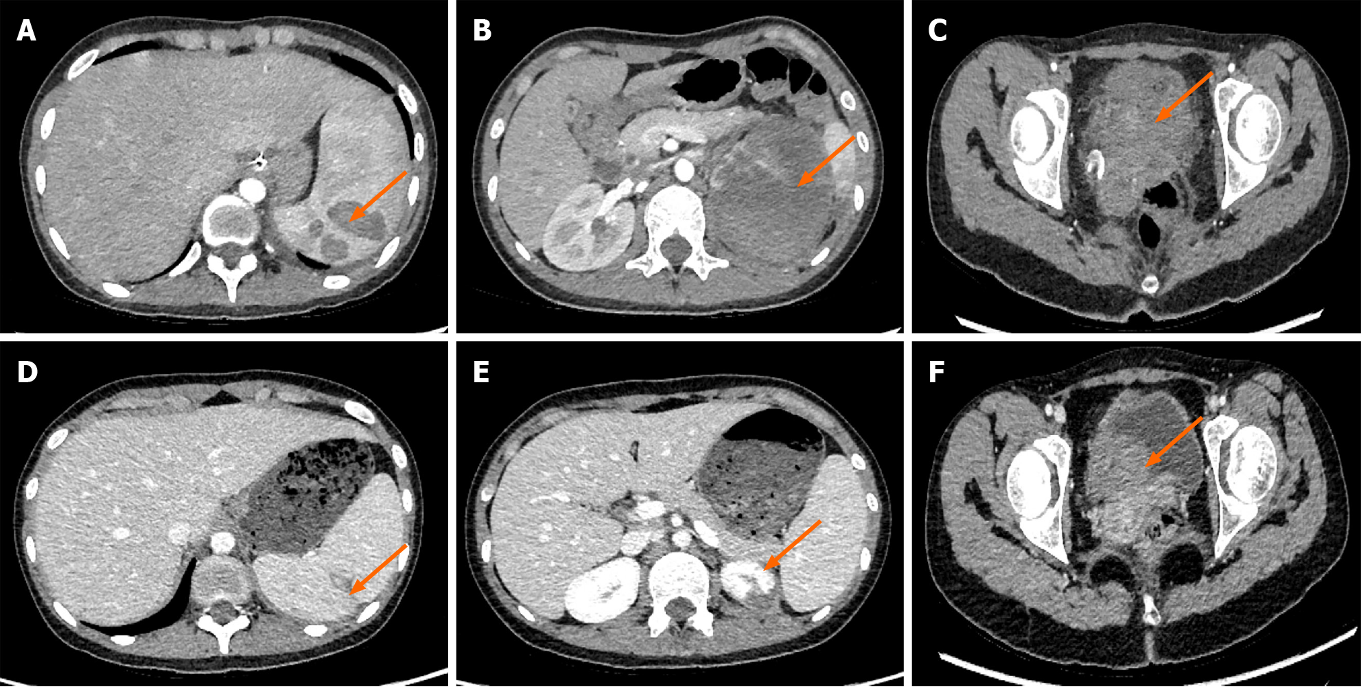
Figure 6 Abdominal computed tomography images of the patient at different stages of treatment.
A: Multiple nodules of spleen were shown on computed tomography (CT) image before chemotherapy (orange arrows, metastatic nodules); B: CT images revealed an almost normal spleen after treatment; C: Large cystic occupying lesion of left renal was shown on CT image before chemotherapy (orange arrows, metastatic cystic occupying lesion); D: CT images revealed an almost normal but smaller left kidney after treatment; E: Large solid occupying lesion of cervix was shown on CT image before chemotherapy (orange arrows, metastatic solid occupying lesion); F: CT images revealed an almost normal cervix after treatment.
- Citation: Huang HQ, Gong FM, Yin RT, Lin XJ. Choriocarcinoma misdiagnosed as cerebral hemangioma: A case report . World J Clin Cases 2021; 9(30): 9174-9181
- URL: https://www.wjgnet.com/2307-8960/full/v9/i30/9174.htm
- DOI: https://dx.doi.org/10.12998/wjcc.v9.i30.9174


