Copyright
©The Author(s) 2021.
World J Clin Cases. Oct 26, 2021; 9(30): 9159-9167
Published online Oct 26, 2021. doi: 10.12998/wjcc.v9.i30.9159
Published online Oct 26, 2021. doi: 10.12998/wjcc.v9.i30.9159
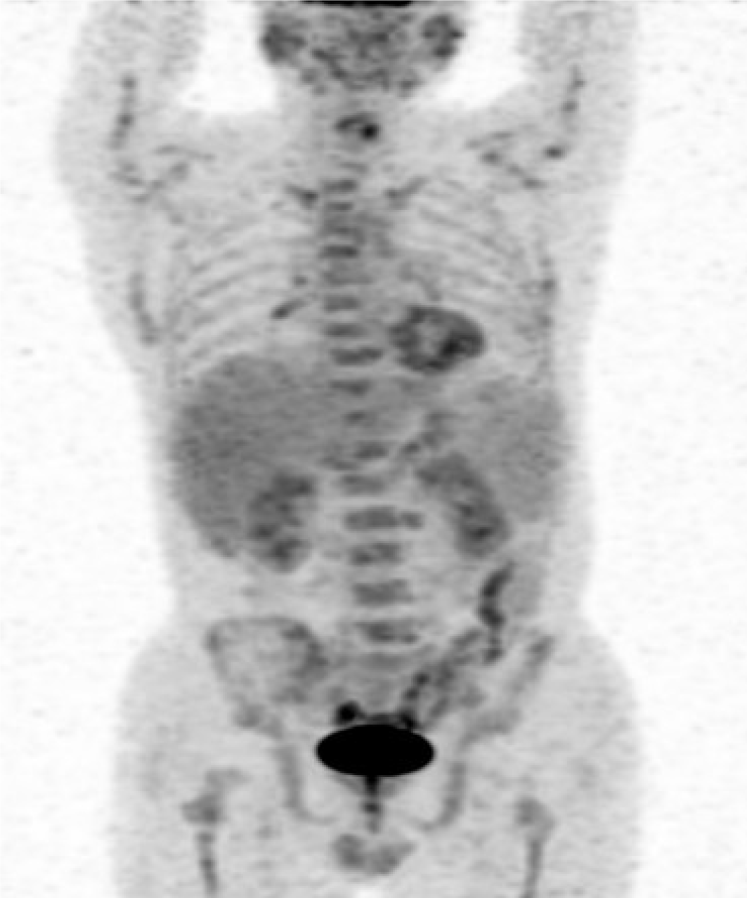
Figure 4 Positron emission tomography-computed tomography scanning in Case 2.
The maximum intensity projection of 18F-fluorodeoxyglucose positron emission tomography-computed tomography revealed that the spleen was enlarged and 18F-fluorodeoxyglucose uptake was normal. Hypermetabolic lesions were detected in bone marrow, bilateral inguinal and bilateral lung hilar lymphadenopathy.
- Citation: Shen J, Wang JS, Xie JL, Nong L, Chen JN, Wang Z. Hemophagocytic lymphohistiocytosis secondary to composite lymphoma: Two case reports. World J Clin Cases 2021; 9(30): 9159-9167
- URL: https://www.wjgnet.com/2307-8960/full/v9/i30/9159.htm
- DOI: https://dx.doi.org/10.12998/wjcc.v9.i30.9159


