Copyright
©The Author(s) 2021.
World J Clin Cases. Oct 26, 2021; 9(30): 9159-9167
Published online Oct 26, 2021. doi: 10.12998/wjcc.v9.i30.9159
Published online Oct 26, 2021. doi: 10.12998/wjcc.v9.i30.9159
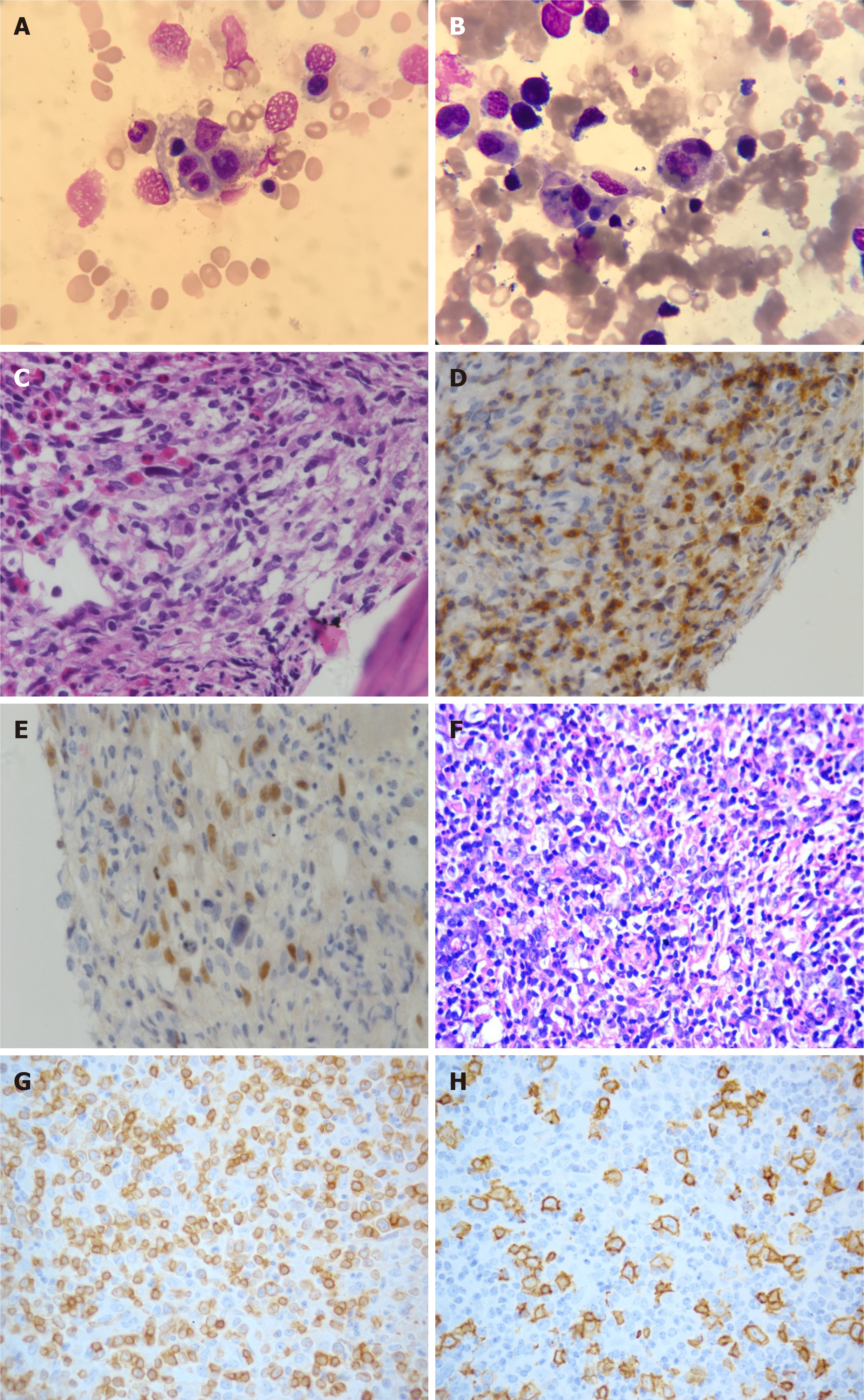
Figure 1 Morphology of bone marrow aspirate smear and biopsy in Case 1.
A and B: Hemophagocytosis (A) and tumor cells (B) (Wright’s stain, × 1000); C: Diffuse small to medium-sized lymphocytes admixed scattered large atypical cells (hematoxylin-eosin stain, × 400); D and E: The tumor cells were positive for CD3 (D) and PAX-5 (E) (immunohistochemistry, IHC × 400); F: Lymph node biopsy sections showed a marked nodular aggregate of medium-sized lymphocytes with scattered large lymphocytes (hematoxylin-eosin stain, × 400); G and H: The tumor cells in lymph nodes were positive for CD3 (G) and CD20 (H) (IHC × 400).
- Citation: Shen J, Wang JS, Xie JL, Nong L, Chen JN, Wang Z. Hemophagocytic lymphohistiocytosis secondary to composite lymphoma: Two case reports. World J Clin Cases 2021; 9(30): 9159-9167
- URL: https://www.wjgnet.com/2307-8960/full/v9/i30/9159.htm
- DOI: https://dx.doi.org/10.12998/wjcc.v9.i30.9159


