Copyright
©The Author(s) 2021.
World J Clin Cases. Oct 26, 2021; 9(30): 9122-9128
Published online Oct 26, 2021. doi: 10.12998/wjcc.v9.i30.9122
Published online Oct 26, 2021. doi: 10.12998/wjcc.v9.i30.9122
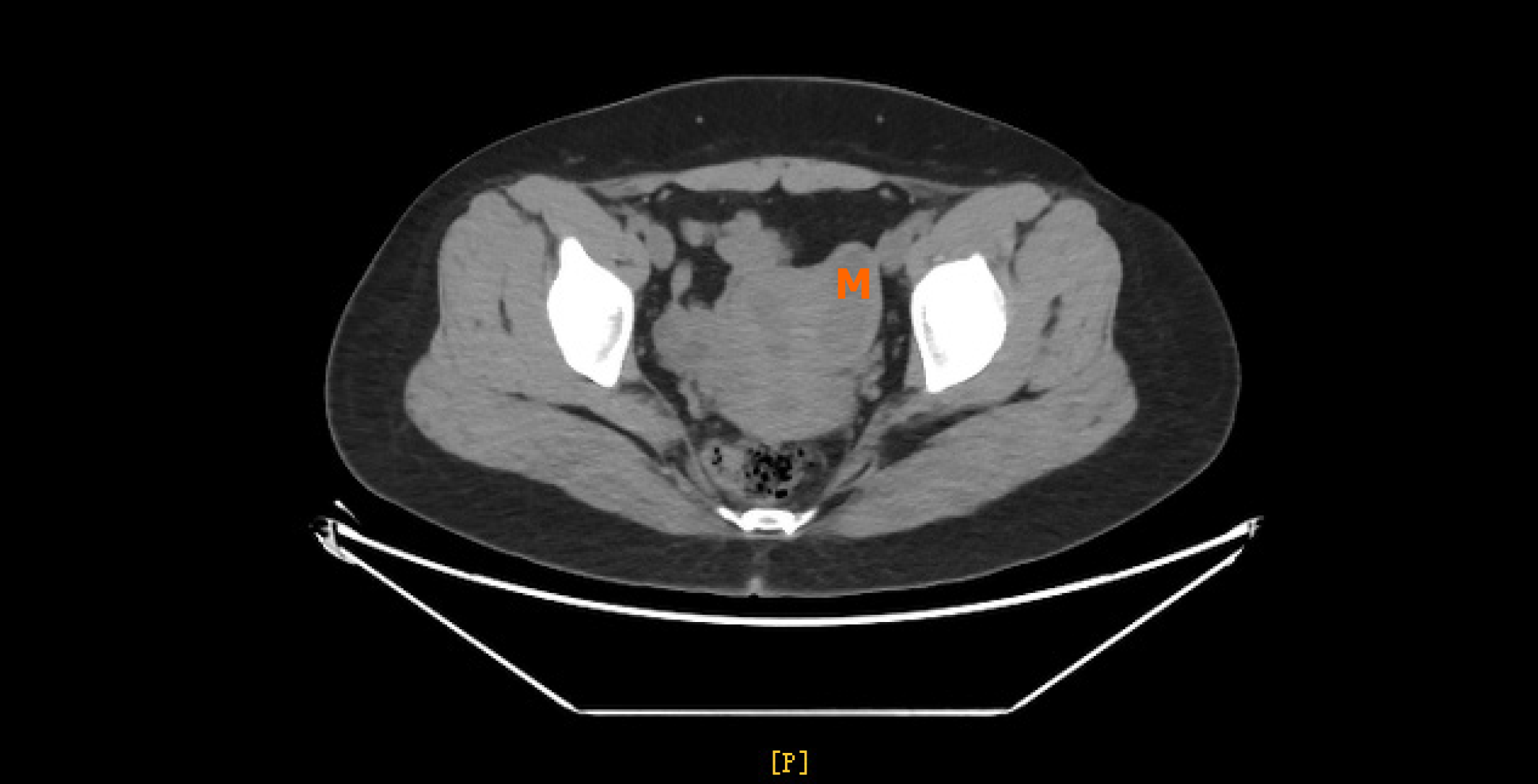
Figure 2 Computed tomography showing a cystic lesion with a size of about 4.
9 cm × 2.0 cm × 2.1 cm on the left side of the pelvis, not clearly demarcated from the left uterine muscle wall. M: Mass.
- Citation: Hu YL, Wang A, Chen J. Diagnosis and laparoscopic excision of accessory cavitated uterine mass in a young woman: A case report. World J Clin Cases 2021; 9(30): 9122-9128
- URL: https://www.wjgnet.com/2307-8960/full/v9/i30/9122.htm
- DOI: https://dx.doi.org/10.12998/wjcc.v9.i30.9122


