Copyright
©The Author(s) 2021.
World J Clin Cases. Oct 26, 2021; 9(30): 8974-8984
Published online Oct 26, 2021. doi: 10.12998/wjcc.v9.i30.8974
Published online Oct 26, 2021. doi: 10.12998/wjcc.v9.i30.8974
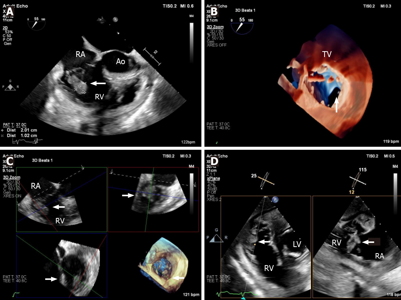
Figure 3 Transesophageal echocardiography in native tricuspid valve endocarditis.
A: Mid-esophageal modified short-axis view showing a large vegetation attached to the native tricuspid valve (arrow); B: 3D transesophageal echocardiography imaging showing the vegetation to arise from posterior tricuspid leaflet; C: 3D multi-planar reconstruction on the vegetation, confirming the location on the posterior leaflet (arrow); D: Transgatric view, showing multi-planar assessment of the vegetation. RA: Right atrium; RV: Right ventricle; LA: Left atrium; LV: Left ventricle; TV: Tricuspid valve.
- Citation: Fava AM, Xu B. Tricuspid valve endocarditis: Cardiovascular imaging evaluation and management. World J Clin Cases 2021; 9(30): 8974-8984
- URL: https://www.wjgnet.com/2307-8960/full/v9/i30/8974.htm
- DOI: https://dx.doi.org/10.12998/wjcc.v9.i30.8974


