Copyright
©The Author(s) 2021.
World J Clin Cases. Oct 26, 2021; 9(30): 8974-8984
Published online Oct 26, 2021. doi: 10.12998/wjcc.v9.i30.8974
Published online Oct 26, 2021. doi: 10.12998/wjcc.v9.i30.8974
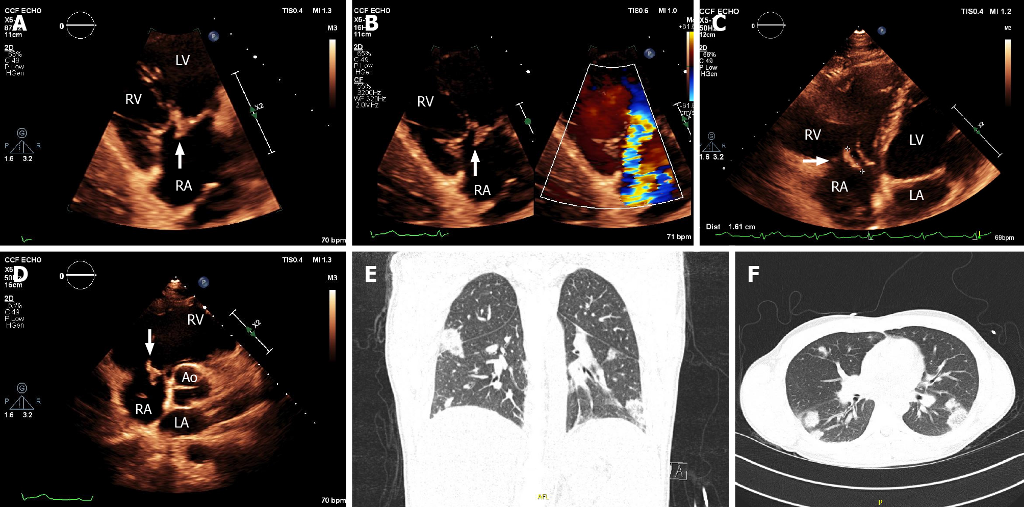
Figure 1 Transthoracic echocardiography and computed tomography imaging assessment of native tricuspid valve endocarditis.
A: Modified apical four-chambers view, focused on the tricuspid valve showing a vegetation attached on tricuspid valve (arrow); B: Color Doppler analysis showing severe tricuspid regurgitation caused by the vegetation; C: Sub-xiphoid view, measuring the vegetation as 1.6 cm; D: Short-axis view, showing the vegetation on the tricuspid valve prolapsing into the right atrium (arrow); E and F: Computed tomography lung demonstrating multiple areas of septic cavitations. RA: Right atrium; RV: Right ventricle; LA: Left atrium; LV: Left ventricle.
- Citation: Fava AM, Xu B. Tricuspid valve endocarditis: Cardiovascular imaging evaluation and management. World J Clin Cases 2021; 9(30): 8974-8984
- URL: https://www.wjgnet.com/2307-8960/full/v9/i30/8974.htm
- DOI: https://dx.doi.org/10.12998/wjcc.v9.i30.8974


