Copyright
©The Author(s) 2021.
World J Clin Cases. Jan 26, 2021; 9(3): 736-747
Published online Jan 26, 2021. doi: 10.12998/wjcc.v9.i3.736
Published online Jan 26, 2021. doi: 10.12998/wjcc.v9.i3.736
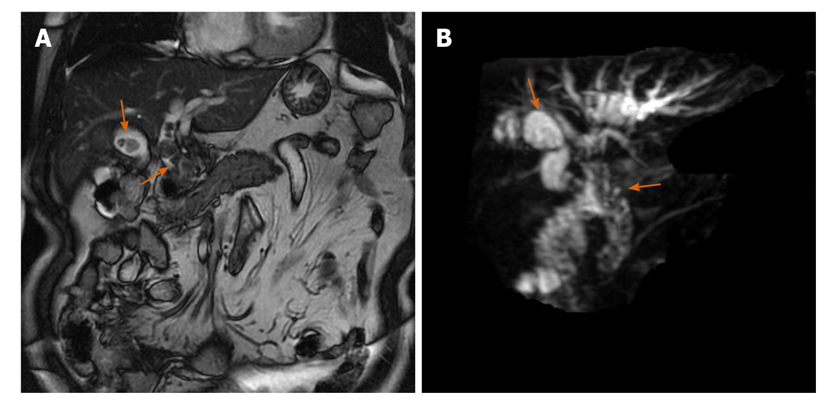
Figure 1 Preoperative magnetic resonance cholangiopancreatography showed gallbladder and common bile duct stones.
The orange arrowpoints to the stone location. A: T2 weighted image sequence; B: Balanced turbo field echo sequence.
- Citation: He YG, Gao MF, Li J, Peng XH, Tang YC, Huang XB, Li YM. Cystic duct dilation through endoscopic retrograde cholangiopancreatography for treatment of gallstones and choledocholithiasis: Six case reports and review of literature. World J Clin Cases 2021; 9(3): 736-747
- URL: https://www.wjgnet.com/2307-8960/full/v9/i3/736.htm
- DOI: https://dx.doi.org/10.12998/wjcc.v9.i3.736


