Copyright
©The Author(s) 2021.
World J Clin Cases. Jan 26, 2021; 9(3): 666-671
Published online Jan 26, 2021. doi: 10.12998/wjcc.v9.i3.666
Published online Jan 26, 2021. doi: 10.12998/wjcc.v9.i3.666
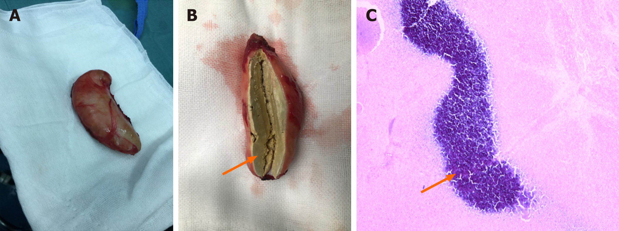
Figure 3 Gross specimen and biopsy of the resected mass.
A: The gross specimen of the mass showing an encapsulated nodule; B: The cut surface of the mass. The arrowhead indicates the necrotic tissue inside; C: The biopsy of the mass. The cystic wall of the mass consists of fibrous tissue. The blue-stained area indicated by the arrowhead is non-structured necrotic tissue.
- Citation: Xie Y, Luo YR, Chen M, Xie YM, Sun CY, Chen Q. Pleural lump after paragonimiasis treated by thoracoscopy: A case report. World J Clin Cases 2021; 9(3): 666-671
- URL: https://www.wjgnet.com/2307-8960/full/v9/i3/666.htm
- DOI: https://dx.doi.org/10.12998/wjcc.v9.i3.666


