Copyright
©The Author(s) 2021.
World J Clin Cases. Oct 16, 2021; 9(29): 8938-8945
Published online Oct 16, 2021. doi: 10.12998/wjcc.v9.i29.8938
Published online Oct 16, 2021. doi: 10.12998/wjcc.v9.i29.8938
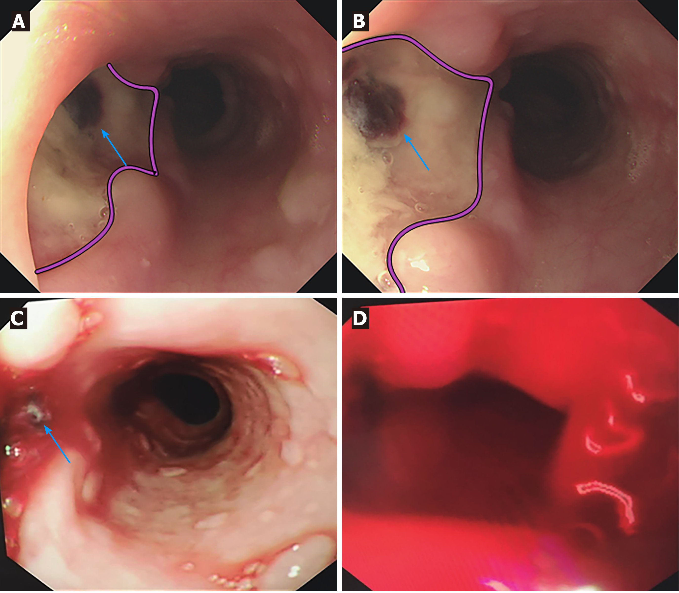
Figure 2 Esophageal endoscopic images of the patient.
A and B: First esophageal endoscopy showed a large deep longitudinal ulceration (2 cm × 1 cm) with an adherent clot (blue arrows) and white coating 25-27 cm below the incisors, corresponding to a fistula (pink lines showed the margin of the ulcer). No bleeding was found; C: Second bedside emergency endoscopy also showed blood clots (blue arrows); D: Second bedside emergency endoscopy showed massive bleeding as well.
- Citation: Wang DQ, Liu M, Fan WJ. Secondary aortoesophageal fistula initially presented with empyema after thoracic aortic stent grafting: A case report. World J Clin Cases 2021; 9(29): 8938-8945
- URL: https://www.wjgnet.com/2307-8960/full/v9/i29/8938.htm
- DOI: https://dx.doi.org/10.12998/wjcc.v9.i29.8938


