Copyright
©The Author(s) 2021.
World J Clin Cases. Oct 16, 2021; 9(29): 8938-8945
Published online Oct 16, 2021. doi: 10.12998/wjcc.v9.i29.8938
Published online Oct 16, 2021. doi: 10.12998/wjcc.v9.i29.8938
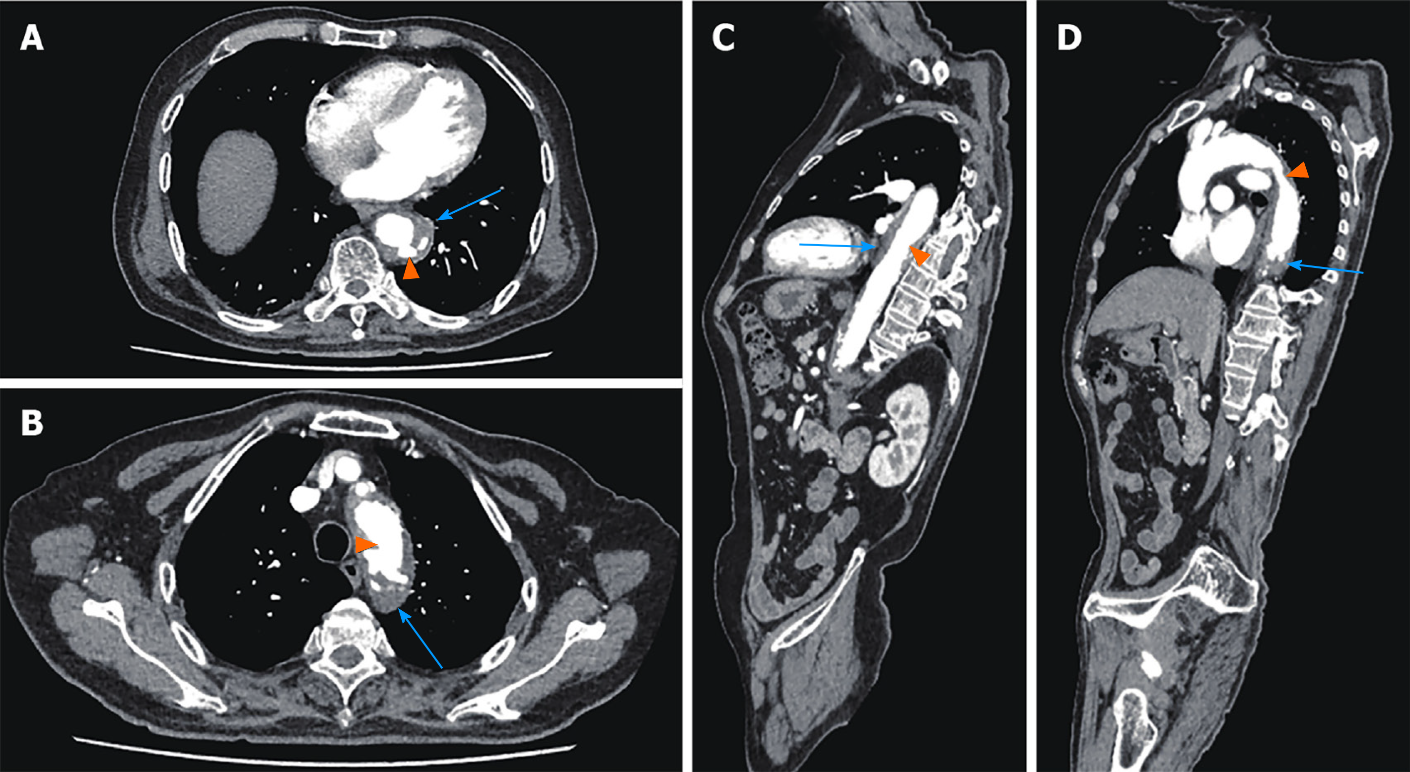
Figure 1 Preoperative thoracic and abdominal aortic computed tomography angiography.
A and B: Axial Computed tomography (CT) angiography showed low-density shadow peri-descending aorta, indicating intramural hemorrhage and hematoma (blue arrows), and multiple penetrating ulcers (orange arrows); C and D: Sagittal CT angiography showed low-density shadow peri-descending aorta, indicating intramural hemorrhage and hematoma (blue arrows), and multiple penetrating ulcers (orange arrows).
- Citation: Wang DQ, Liu M, Fan WJ. Secondary aortoesophageal fistula initially presented with empyema after thoracic aortic stent grafting: A case report. World J Clin Cases 2021; 9(29): 8938-8945
- URL: https://www.wjgnet.com/2307-8960/full/v9/i29/8938.htm
- DOI: https://dx.doi.org/10.12998/wjcc.v9.i29.8938


