Copyright
©The Author(s) 2021.
World J Clin Cases. Oct 16, 2021; 9(29): 8901-8905
Published online Oct 16, 2021. doi: 10.12998/wjcc.v9.i29.8901
Published online Oct 16, 2021. doi: 10.12998/wjcc.v9.i29.8901
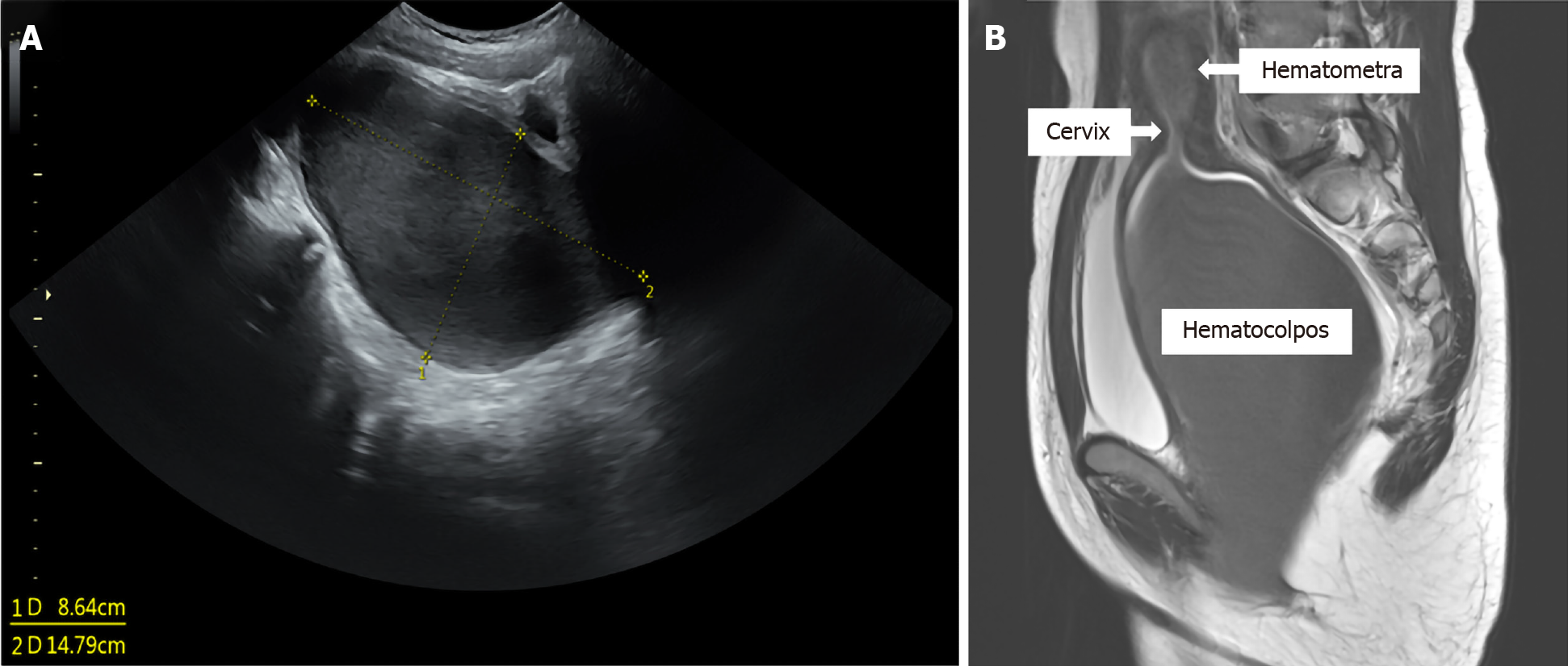
Figure 1 Radiological images.
A: Transabdominal ultrasound shows a hypoechoic cystic mass below the bladder (14.79 cm × 8.64 cm); B: Pelvic magnetic resonance imaging shows marked distention of the uterus and vagina with blood clots.
- Citation: Jang E, So KA, Kim B, Lee AJ, Kim NR, Yang EJ, Shim SH, Lee SJ, Kim TJ. Delayed diagnosis of imperforate hymen with huge hematocolpometra: A case report. World J Clin Cases 2021; 9(29): 8901-8905
- URL: https://www.wjgnet.com/2307-8960/full/v9/i29/8901.htm
- DOI: https://dx.doi.org/10.12998/wjcc.v9.i29.8901


