Copyright
©The Author(s) 2021.
World J Clin Cases. Oct 16, 2021; 9(29): 8888-8893
Published online Oct 16, 2021. doi: 10.12998/wjcc.v9.i29.8888
Published online Oct 16, 2021. doi: 10.12998/wjcc.v9.i29.8888
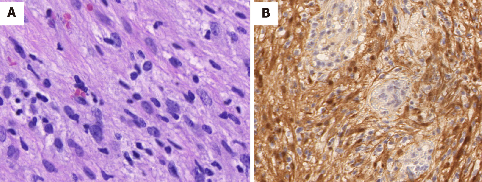
Figure 3 Pathological images of tumor cells.
A: A histologic section from the tumor shows that the tumor cells were spindled with elongated palisading nuclei (hematoxylin–eosin stain, 400 ×); B: The tumor cells showed diffuse and positive S-100 staining by immunohistochemistry (streptavidin peroxidase method, 200 ×).
- Citation: Wu L, Sha MC, Wu XL, Bi J, Chen ZM, Wang YS. Primary intratracheal neurilemmoma in a 10-year-old girl: A case report. World J Clin Cases 2021; 9(29): 8888-8893
- URL: https://www.wjgnet.com/2307-8960/full/v9/i29/8888.htm
- DOI: https://dx.doi.org/10.12998/wjcc.v9.i29.8888


