Copyright
©The Author(s) 2021.
World J Clin Cases. Oct 16, 2021; 9(29): 8879-8887
Published online Oct 16, 2021. doi: 10.12998/wjcc.v9.i29.8879
Published online Oct 16, 2021. doi: 10.12998/wjcc.v9.i29.8879
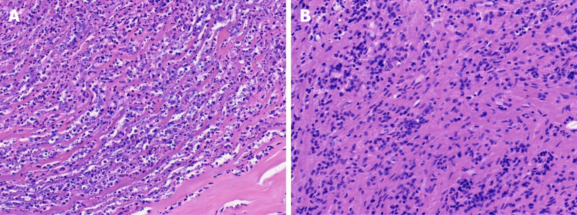
Figure 4 Histological staining of lumbar 1/2 focal tissue.
A: Haematoxylin-eosin staining (40 × magnification) showing acute purulent inflammation and inflammatory necrosis; B: Haematoxylin-eosin staining (magnification × 40) showing chronic inflammatory cell infiltration.
- Citation: Tan YZ, Yuan T, Tan L, Tian YQ, Long YZ. Lumbar infection caused by Mycobacterium paragordonae: A case report. World J Clin Cases 2021; 9(29): 8879-8887
- URL: https://www.wjgnet.com/2307-8960/full/v9/i29/8879.htm
- DOI: https://dx.doi.org/10.12998/wjcc.v9.i29.8879


