Copyright
©The Author(s) 2021.
World J Clin Cases. Oct 16, 2021; 9(29): 8871-8878
Published online Oct 16, 2021. doi: 10.12998/wjcc.v9.i29.8871
Published online Oct 16, 2021. doi: 10.12998/wjcc.v9.i29.8871
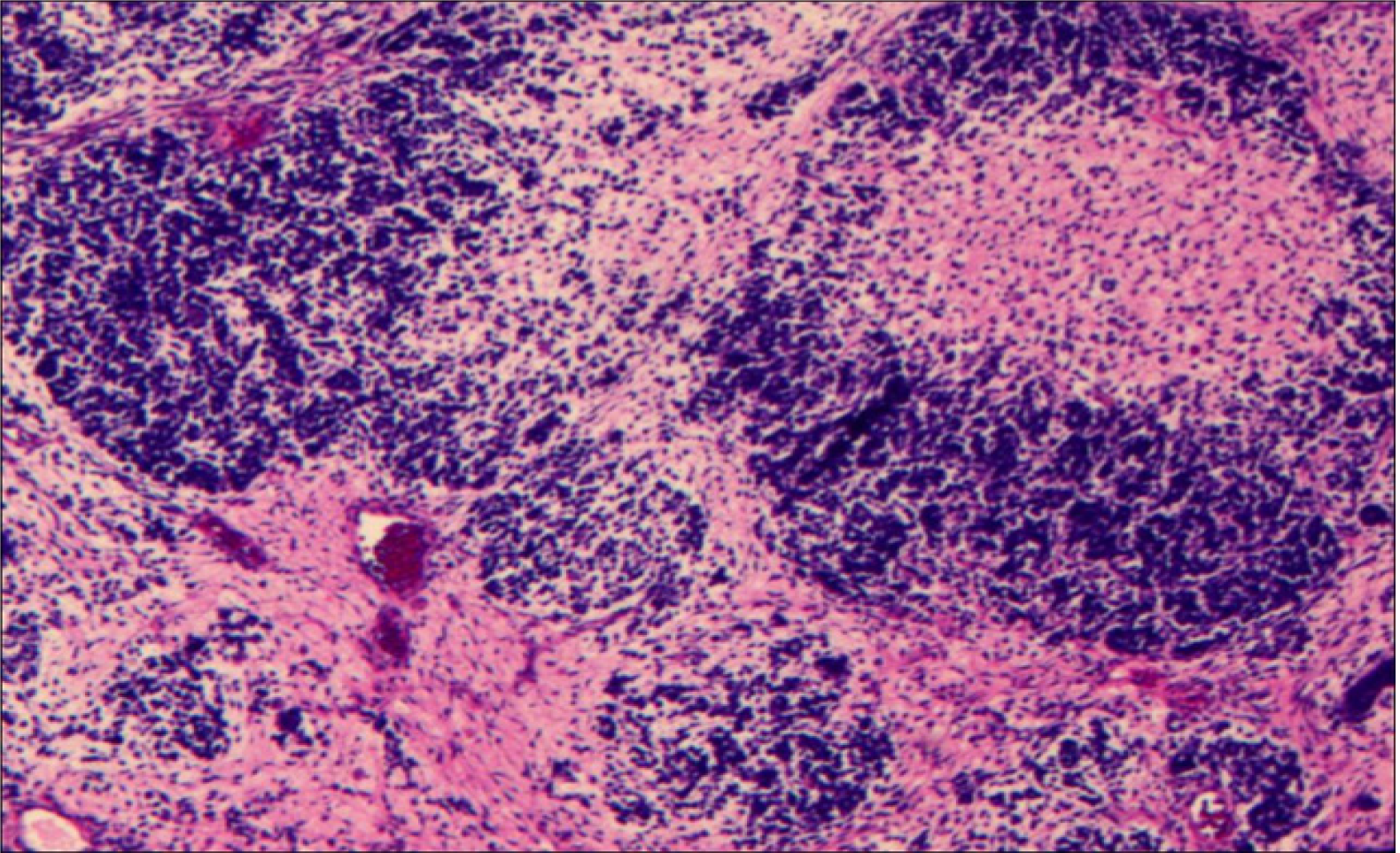
Figure 3 Pathological examination of the tumor tissue.
Hematoxylin–eosin staining of the tumor cells showed diffuse growth with cellular atypia and mitotic figures. Image at 40 × magnification.
- Citation: Wang YY, Li ML, Zhang ZY, Ding JW, Xiao LF, Li WC, Wang L, Sun T. Primary intracranial synovial sarcoma with hemorrhage: A case report. World J Clin Cases 2021; 9(29): 8871-8878
- URL: https://www.wjgnet.com/2307-8960/full/v9/i29/8871.htm
- DOI: https://dx.doi.org/10.12998/wjcc.v9.i29.8871


