Copyright
©The Author(s) 2021.
World J Clin Cases. Oct 16, 2021; 9(29): 8864-8870
Published online Oct 16, 2021. doi: 10.12998/wjcc.v9.i29.8864
Published online Oct 16, 2021. doi: 10.12998/wjcc.v9.i29.8864
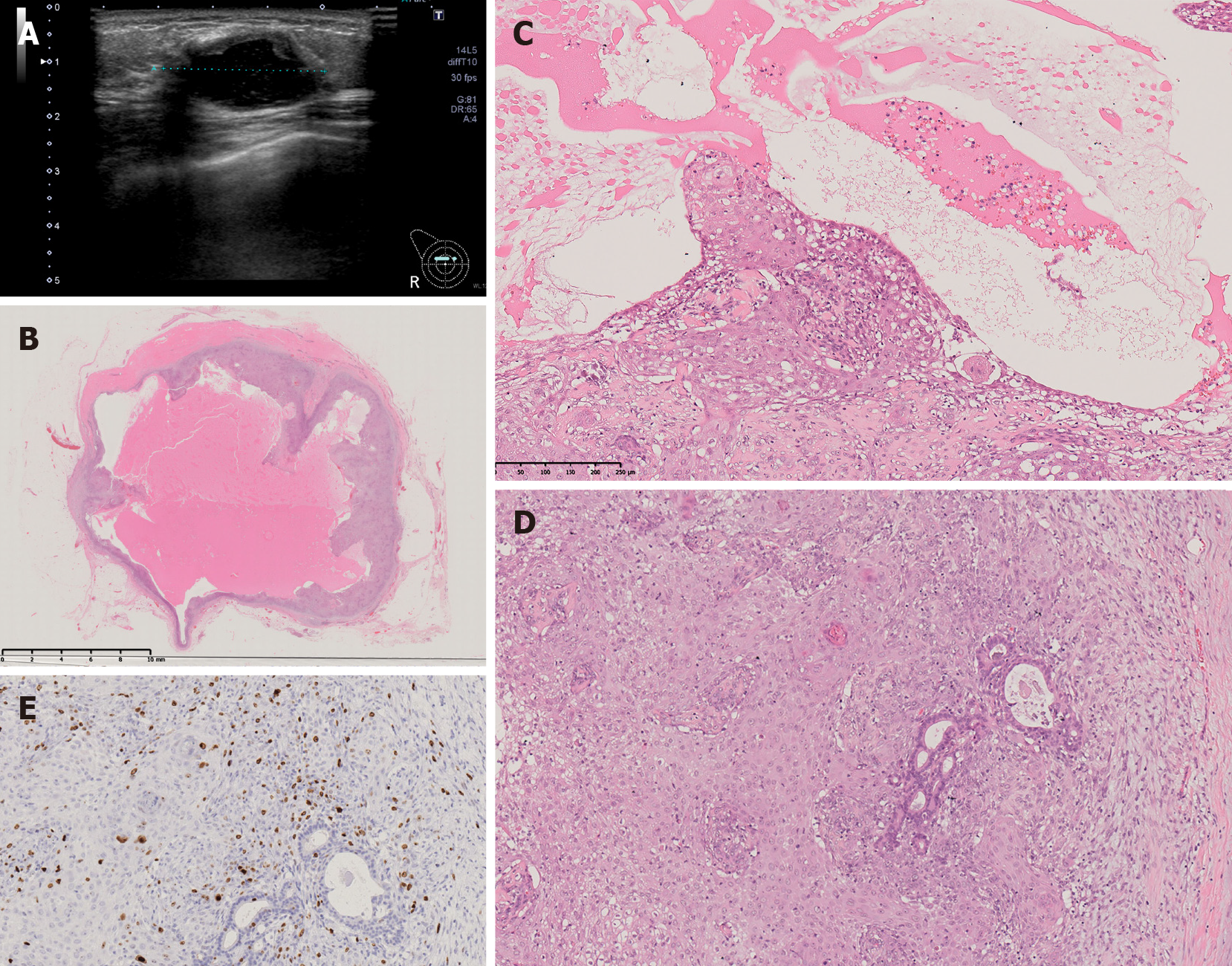
Figure 2 Imaging and pathology findings at first recurrence.
A: Ultrasonography showed a well-defined, oval, low-isoechoic mass; B: The tumor was a cystic lesion, and the cystic wall had nodules or irregular thickening. The cysts contained mucus (× 32 magnification); C and D: Epithelium with squamous metaplasia growing into the lumenal side (C: × 100 magnification, lumen side) and (D: × 100, membrane side). Dense growth of spindle-shaped myoepithelium is found on the side of the membrane. The myoepithelium spreads underneath the existing glandular epithelium. Tumor cells show prominent nuclear atypia and high mitotic counts; E: The Ki 67 hot spot is 57.1% (× 100 magnification, Ki67).
- Citation: Oda G, Nakagawa T, Mori M, Fujioka T, Onishi I. Adenomyoepithelioma of the breast with malignant transformation and repeated local recurrence: A case report. World J Clin Cases 2021; 9(29): 8864-8870
- URL: https://www.wjgnet.com/2307-8960/full/v9/i29/8864.htm
- DOI: https://dx.doi.org/10.12998/wjcc.v9.i29.8864


