Copyright
©The Author(s) 2021.
World J Clin Cases. Oct 16, 2021; 9(29): 8858-8863
Published online Oct 16, 2021. doi: 10.12998/wjcc.v9.i29.8858
Published online Oct 16, 2021. doi: 10.12998/wjcc.v9.i29.8858
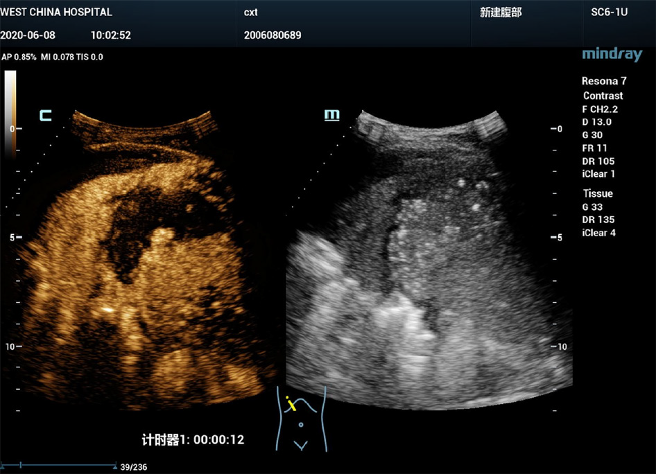
Figure 2
First-time intravenous contrast-enhanced ultrasound showed that the right subphrenic mass (6.
0 cm × 3.0 cm) presented a large-scale non-enhancement area with slightly enhanced septa inside.
- Citation: Qiu TT, Fu R, Luo Y, Ling WW. Diagnosis of upper gastrointestinal perforation complicated with fistula formation and subphrenic abscess by contrast-enhanced ultrasound: A case report. World J Clin Cases 2021; 9(29): 8858-8863
- URL: https://www.wjgnet.com/2307-8960/full/v9/i29/8858.htm
- DOI: https://dx.doi.org/10.12998/wjcc.v9.i29.8858


