Copyright
©The Author(s) 2021.
World J Clin Cases. Oct 16, 2021; 9(29): 8839-8845
Published online Oct 16, 2021. doi: 10.12998/wjcc.v9.i29.8839
Published online Oct 16, 2021. doi: 10.12998/wjcc.v9.i29.8839
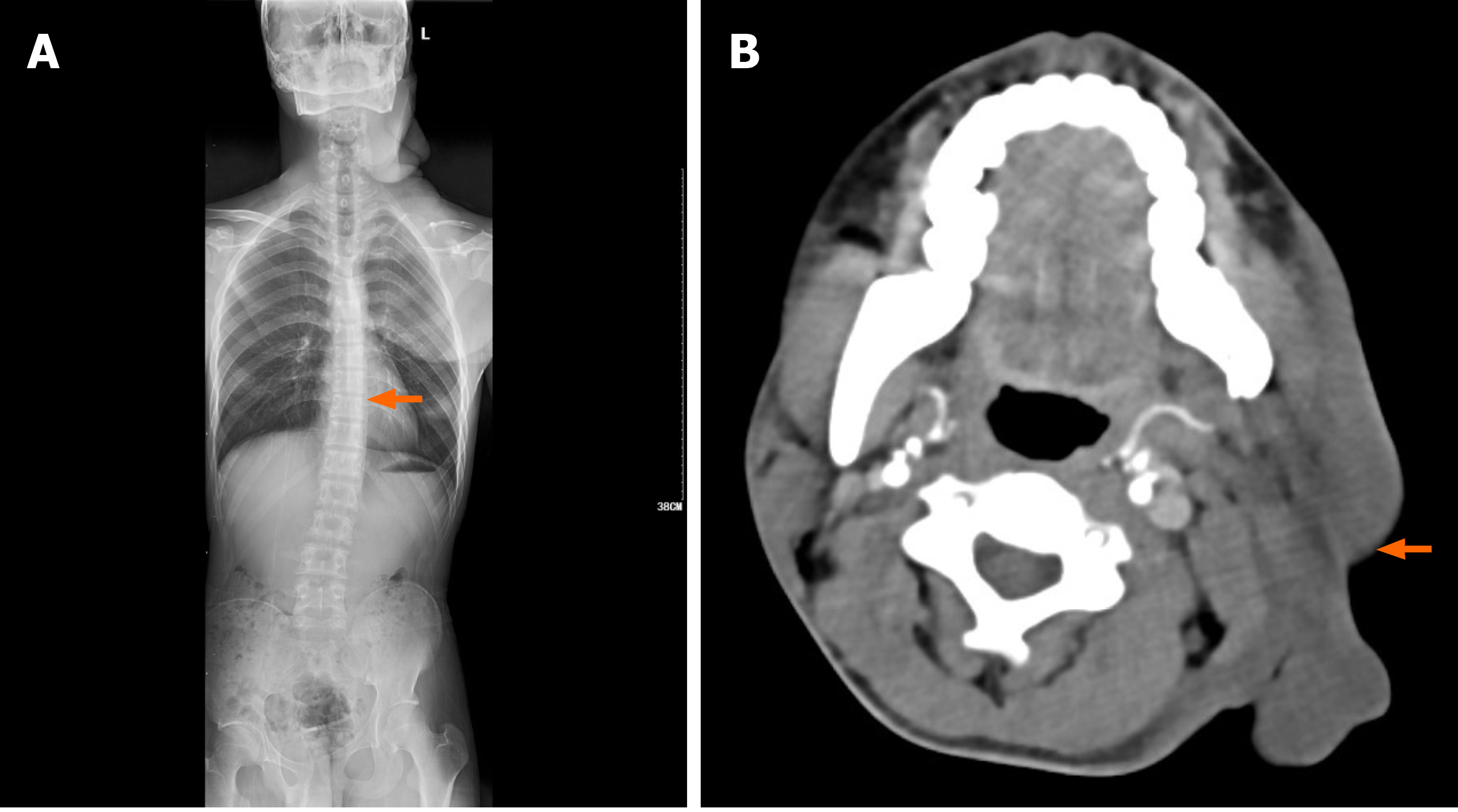
Figure 2 Imaging examination.
A: Digital radiography shows bone changes in the neck, upper chest wall, and surrounding the left shoulder. Scoliosis is also visible; B: Computed tomography (axial section) show soft-tissue changes in the left maxillofacial area.
- Citation: Mu X, Zhang HY, Shen YH, Yang HY. Familial left cervical neurofibromatosis 1 with scoliosis: A case report. World J Clin Cases 2021; 9(29): 8839-8845
- URL: https://www.wjgnet.com/2307-8960/full/v9/i29/8839.htm
- DOI: https://dx.doi.org/10.12998/wjcc.v9.i29.8839


