Copyright
©The Author(s) 2021.
World J Clin Cases. Oct 16, 2021; 9(29): 8831-8838
Published online Oct 16, 2021. doi: 10.12998/wjcc.v9.i29.8831
Published online Oct 16, 2021. doi: 10.12998/wjcc.v9.i29.8831
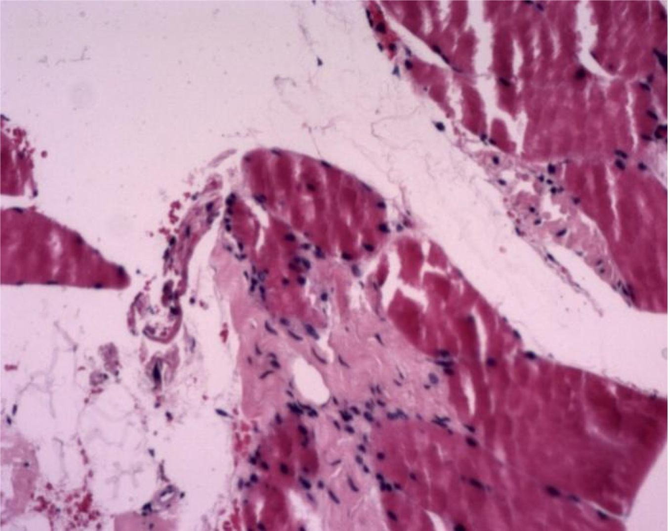
Figure 3 Histopathological examination.
Paraffin section specimens were taken from the right lower limb muscles, stained with histopathological examination, and examined at magnification (100 ×). Skeletal muscles were visible in the broken tissues, some of the horizontal stripes were unclear, and there was proliferation of fibrous tissue and small blood vessels.
- Citation: Song Y, Zhang N, Yu Y. Diagnosis and treatment of eosinophilic fasciitis: Report of two cases. World J Clin Cases 2021; 9(29): 8831-8838
- URL: https://www.wjgnet.com/2307-8960/full/v9/i29/8831.htm
- DOI: https://dx.doi.org/10.12998/wjcc.v9.i29.8831


