Copyright
©The Author(s) 2021.
World J Clin Cases. Oct 16, 2021; 9(29): 8804-8811
Published online Oct 16, 2021. doi: 10.12998/wjcc.v9.i29.8804
Published online Oct 16, 2021. doi: 10.12998/wjcc.v9.i29.8804
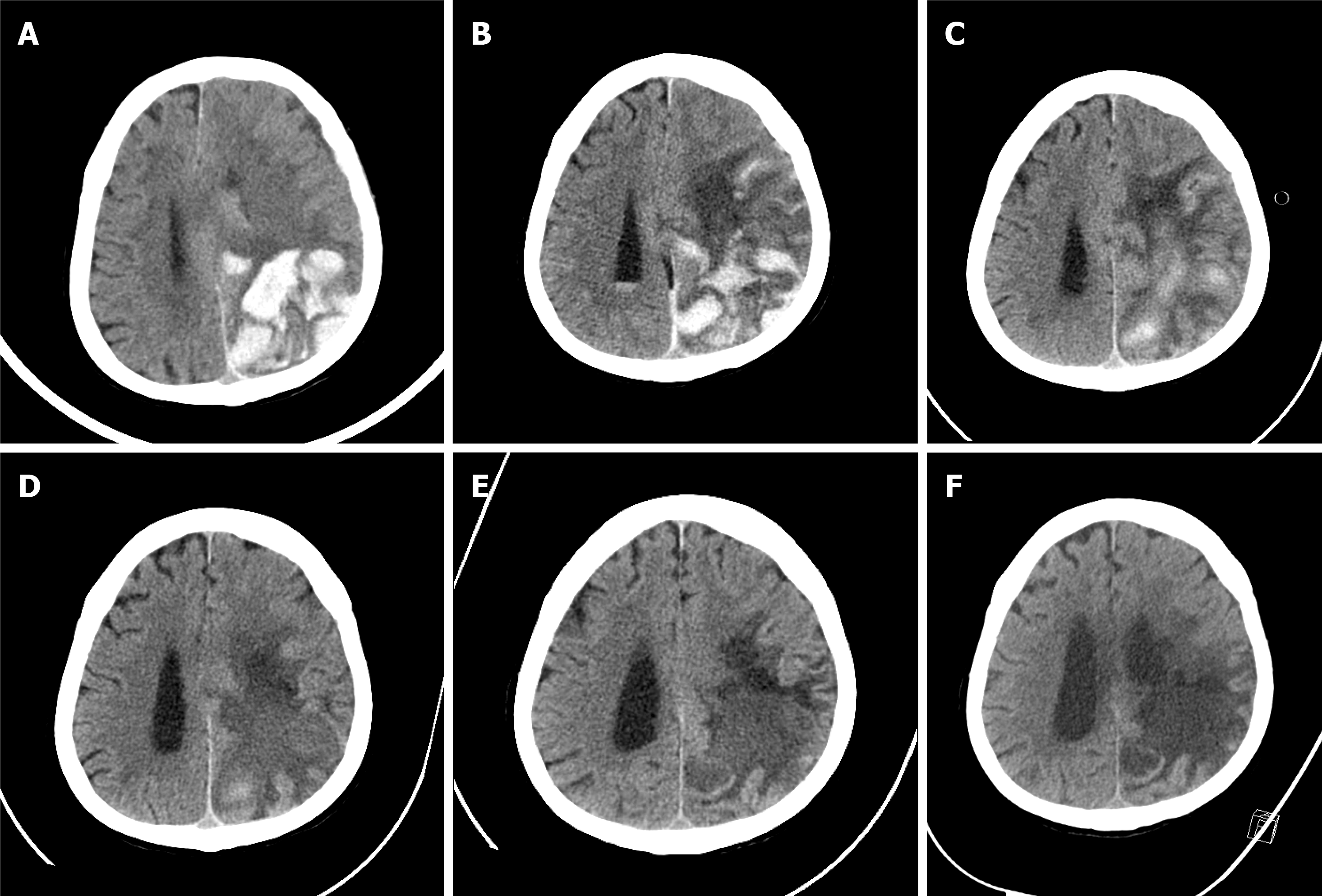
Figure 1 Change in brain computed tomography during hospitalization.
A: Brain computed tomography (CT) at admission. Intracranial hemorrhage in the parieto-occipital lobe was confirmed, and midline shift was observed due to brain edema; B: Brain CT on the 8th hospital day. Intracranial hemorrhage persists and concomitant Intra-ventricular hemorrhage (IVH) is confirmed; C: Brain CT on the 17th hospital day. Intracranial hemorrhage has begun to resolve and improvement of IVH is shown; D: Brain CT on the 28th hospital day. Intracranial hemorrhage shows ongoing resolution, and brain edema has also decreased; E: Brain CT on the 57th hospital day. Improvement in brain edema has resulted in dilatation of the ventricle and resolution of midline shift; F: Brain CT on the 82nd hospital day. Encephalomalacic change in the left parietal lobe is confirmed, and there is no evidence of new intracranial hemorrhage.
- Citation: Kim HS, Lee JY, Jung JW, Lee KH, Kim MJ, Park SB. Is mannitol combined with furosemide a new treatment for refractory lymphedema? A case report. World J Clin Cases 2021; 9(29): 8804-8811
- URL: https://www.wjgnet.com/2307-8960/full/v9/i29/8804.htm
- DOI: https://dx.doi.org/10.12998/wjcc.v9.i29.8804


