Copyright
©The Author(s) 2021.
World J Clin Cases. Oct 16, 2021; 9(29): 8773-8781
Published online Oct 16, 2021. doi: 10.12998/wjcc.v9.i29.8773
Published online Oct 16, 2021. doi: 10.12998/wjcc.v9.i29.8773
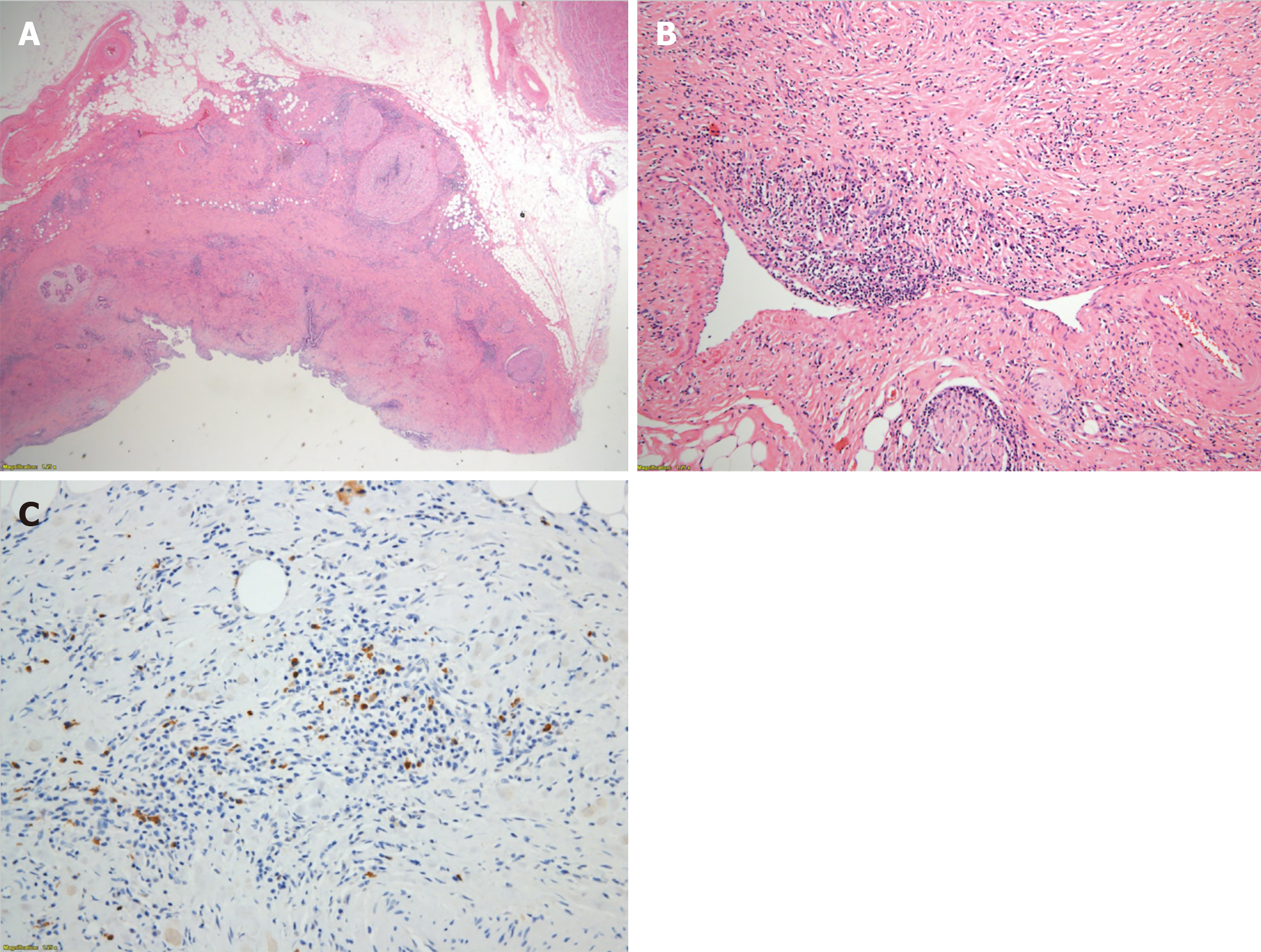
Figure 4 Histopathological examinations.
A: The thickened bile duct wall demonstrates a marked sclerosis with diffuse lymphoplasmacytes and some eosinophils infiltration; B: Obliterative phlebitis is apparent; C: Numerous IgG4+ cells are detected by immunohistochemical staining.
- Citation: Song S, Jo S. Isolated mass-forming IgG4-related sclerosing cholangitis masquerading as extrahepatic cholangiocarcinoma: A case report. World J Clin Cases 2021; 9(29): 8773-8781
- URL: https://www.wjgnet.com/2307-8960/full/v9/i29/8773.htm
- DOI: https://dx.doi.org/10.12998/wjcc.v9.i29.8773


