Copyright
©The Author(s) 2021.
World J Clin Cases. Oct 6, 2021; 9(28): 8616-8626
Published online Oct 6, 2021. doi: 10.12998/wjcc.v9.i28.8616
Published online Oct 6, 2021. doi: 10.12998/wjcc.v9.i28.8616
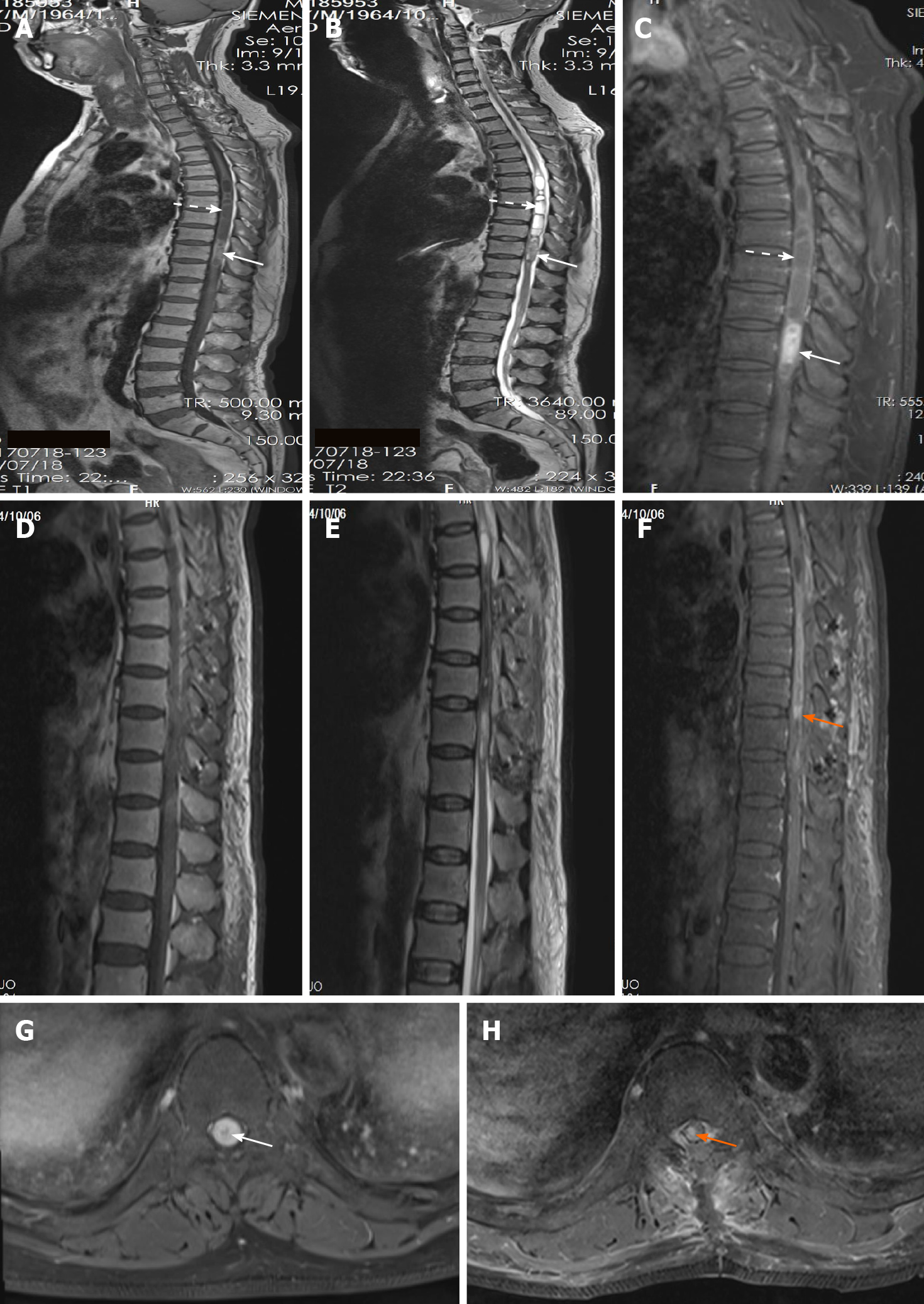
Figure 1 Preoperative and postoperative magnetic resonance imaging.
A: Sagittal T1-weighted image (TIWI); B: Sagittal T2-weighted image (T2WI); C: Sagittal TIWI with gadolinium enhancement; D: Sagittal TIWI; E: Sagittal T2WI; F: Sagittal TIWI with gadolinium enhancement; G: Axial TIWI with gadolinium enhancement at the T10 level; H: Axial TIWI with gadolinium enhancement at the T10 level. A-C and G: Intramedullary melanocytoma (white solid arrows) and syringomyelia (white dashed arrows) were located at the T9-T10 level and T5-T8 level, respectively; D-F and H: An approximate gross total resection of the intramedullary melanocytoma was achieved in spite of tiny residual (orange arrow).
- Citation: Liu ZQ, Liu C, Fu JX, He YQ, Wang Y, Huang TX. Primary intramedullary melanocytoma presenting with lower limbs, defecation, and erectile dysfunction: A case report and review of the literature. World J Clin Cases 2021; 9(28): 8616-8626
- URL: https://www.wjgnet.com/2307-8960/full/v9/i28/8616.htm
- DOI: https://dx.doi.org/10.12998/wjcc.v9.i28.8616


