Copyright
©The Author(s) 2021.
World J Clin Cases. Oct 6, 2021; 9(28): 8518-8523
Published online Oct 6, 2021. doi: 10.12998/wjcc.v9.i28.8518
Published online Oct 6, 2021. doi: 10.12998/wjcc.v9.i28.8518
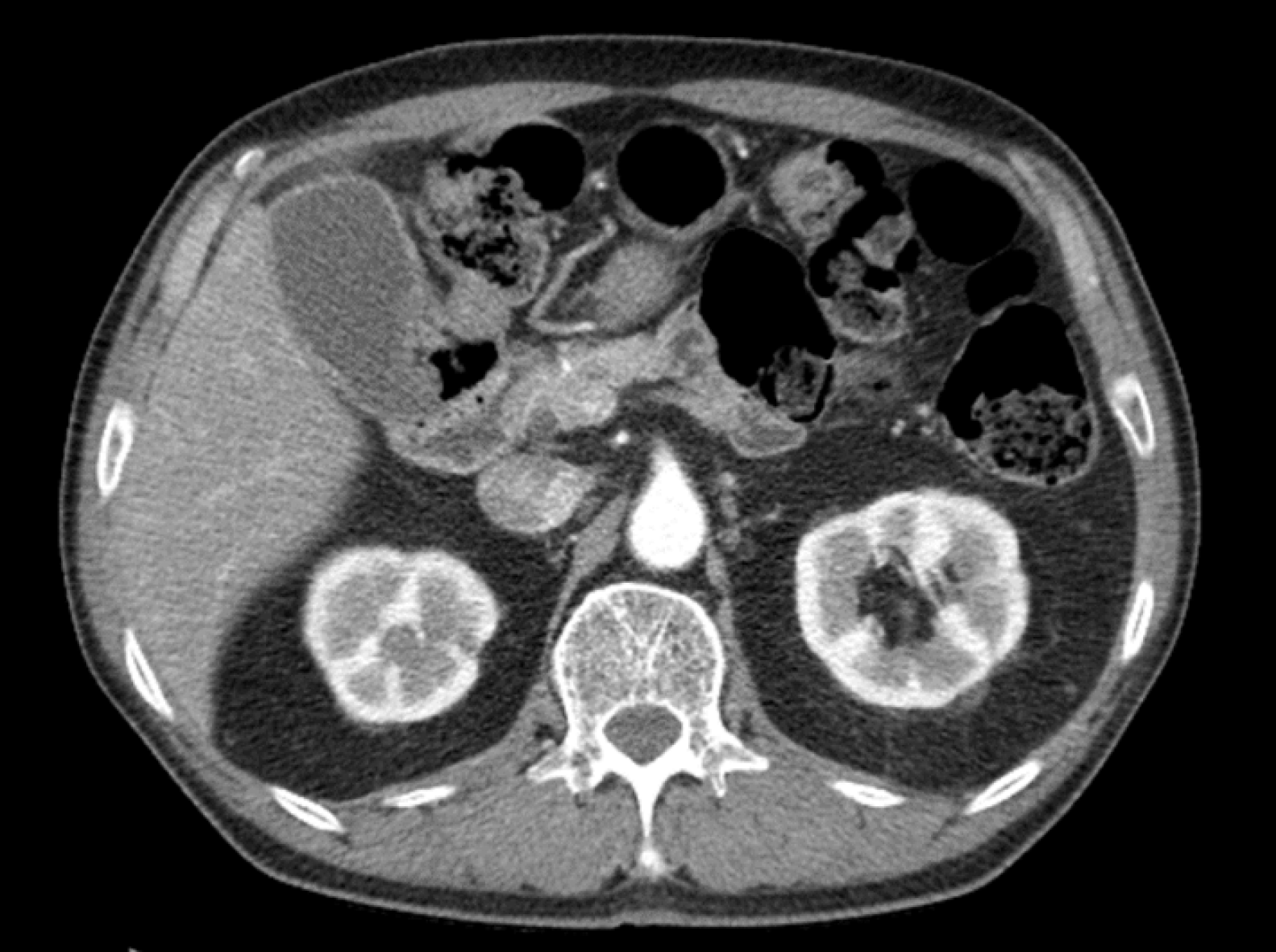
Figure 3 Follow-up computed tomography scan after a 1-month interval.
The findings showed an improved hematoma and a distended gallbladder with mild edematous wall thickening. No gallbladder stone was found.
- Citation: Jang H, Park CH, Park Y, Jeong E, Lee N, Kim J, Jo Y. Spontaneous resolution of gallbladder hematoma in blunt traumatic injury: A case report. World J Clin Cases 2021; 9(28): 8518-8523
- URL: https://www.wjgnet.com/2307-8960/full/v9/i28/8518.htm
- DOI: https://dx.doi.org/10.12998/wjcc.v9.i28.8518


