Copyright
©The Author(s) 2021.
World J Clin Cases. Oct 6, 2021; 9(28): 8518-8523
Published online Oct 6, 2021. doi: 10.12998/wjcc.v9.i28.8518
Published online Oct 6, 2021. doi: 10.12998/wjcc.v9.i28.8518
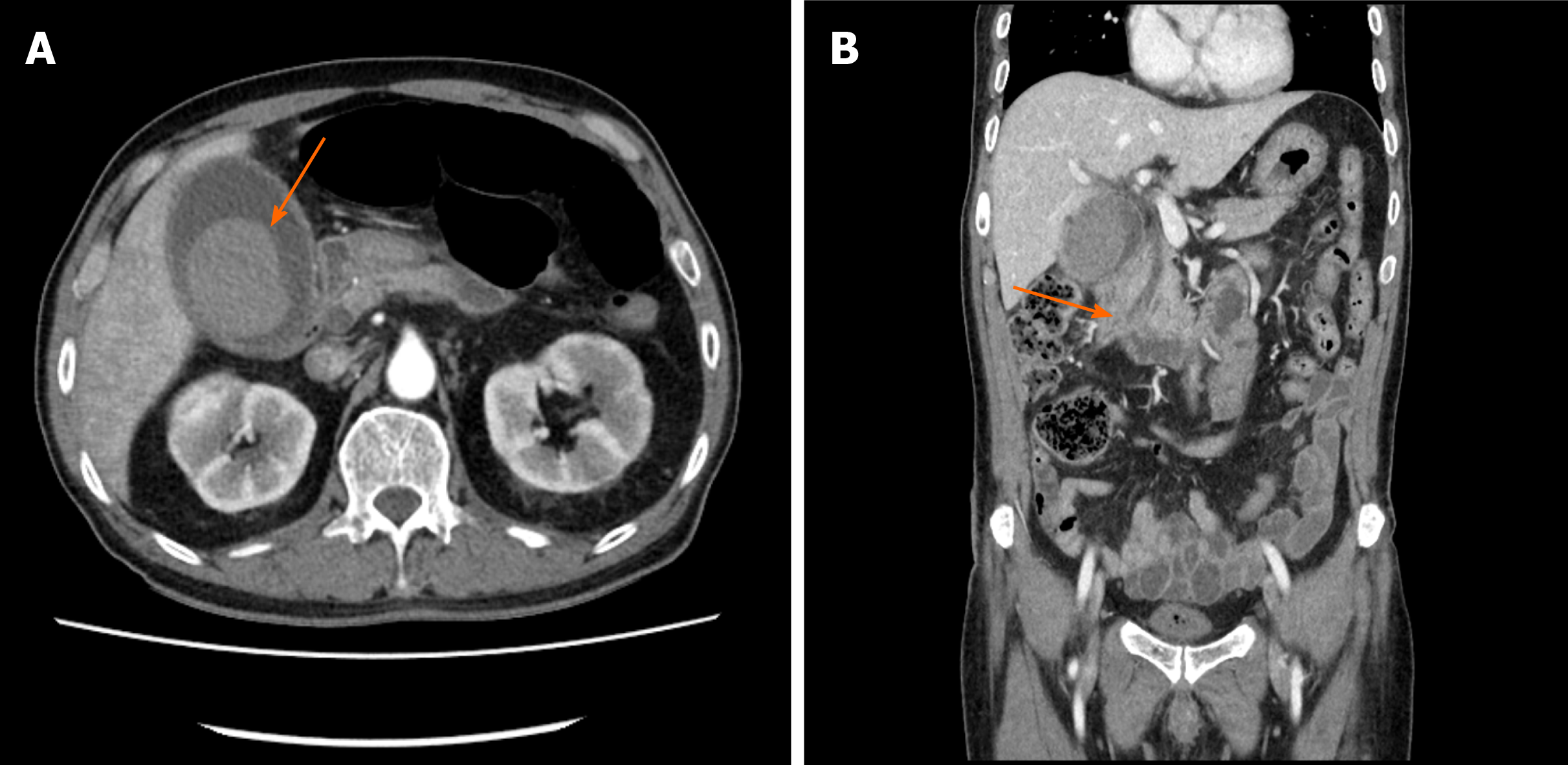
Figure 1 A 55 mm × 40 mm hematoma was seen in the initial computed tomographic scan.
A: The Hounsfield unit values of the gallbladder stone-like lesions ranged from 60 to 67, and no gallbladder wall defect lesion was found (arrow); B: Dilatations of the intrahepatic and common bile ducts are seen (arrow), and there was a suspicion of distal common bile duct obstruction.
- Citation: Jang H, Park CH, Park Y, Jeong E, Lee N, Kim J, Jo Y. Spontaneous resolution of gallbladder hematoma in blunt traumatic injury: A case report. World J Clin Cases 2021; 9(28): 8518-8523
- URL: https://www.wjgnet.com/2307-8960/full/v9/i28/8518.htm
- DOI: https://dx.doi.org/10.12998/wjcc.v9.i28.8518


