Copyright
©The Author(s) 2021.
World J Clin Cases. Oct 6, 2021; 9(28): 8476-8481
Published online Oct 6, 2021. doi: 10.12998/wjcc.v9.i28.8476
Published online Oct 6, 2021. doi: 10.12998/wjcc.v9.i28.8476
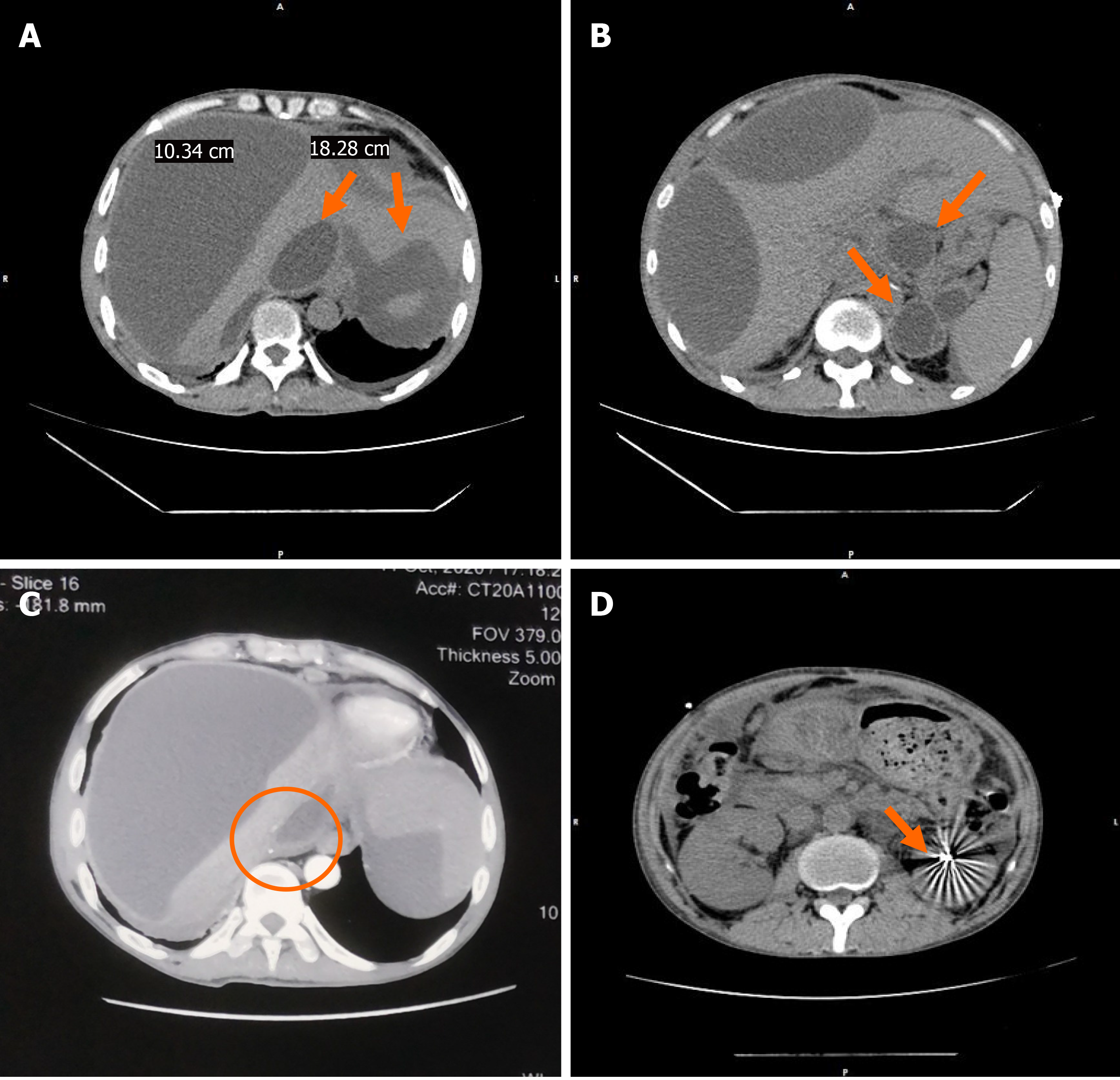
Figure 2 Computed tomography image before percutaneous drainage.
A: Multiple cystic space in the liver (the size of the largest cyst was 18.28 cm × 10.34 cm), and fluid around the spleen; B: Multiple pseudocysts in the pancreatic body and tail; C: Enhanced computed tomography scan showing compressed inferior vena cava; D: High-density shadow after left kidney surgery.
- Citation: Zhu G, Peng YS, Fang C, Yang XL, Li B. Percutaneous drainage in the treatment of intrahepatic pancreatic pseudocyst with Budd-Chiari syndrome: A case report . World J Clin Cases 2021; 9(28): 8476-8481
- URL: https://www.wjgnet.com/2307-8960/full/v9/i28/8476.htm
- DOI: https://dx.doi.org/10.12998/wjcc.v9.i28.8476


