Copyright
©The Author(s) 2021.
World J Clin Cases. Sep 26, 2021; 9(27): 8199-8206
Published online Sep 26, 2021. doi: 10.12998/wjcc.v9.i27.8199
Published online Sep 26, 2021. doi: 10.12998/wjcc.v9.i27.8199
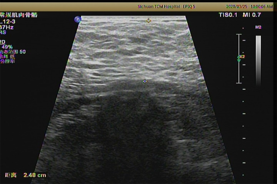
Figure 3 Ultrasound image of a neck mass in the patient.
The subcutaneous fatty masses are seen in the cervical-supraclavicular and occipital regions, being significantly enhanced on both sides, with the thickness of 2.48 cm and unclear borders.
- Citation: Wu L, Jiang T, Zhang Y, Tang AQ, Wu LH, Liu Y, Li MQ, Zhao LB. Madelung’s disease with alcoholic liver disease and acute kidney injury: A case report. World J Clin Cases 2021; 9(27): 8199-8206
- URL: https://www.wjgnet.com/2307-8960/full/v9/i27/8199.htm
- DOI: https://dx.doi.org/10.12998/wjcc.v9.i27.8199


