Copyright
©The Author(s) 2021.
World J Clin Cases. Sep 26, 2021; 9(27): 7963-7972
Published online Sep 26, 2021. doi: 10.12998/wjcc.v9.i27.7963
Published online Sep 26, 2021. doi: 10.12998/wjcc.v9.i27.7963
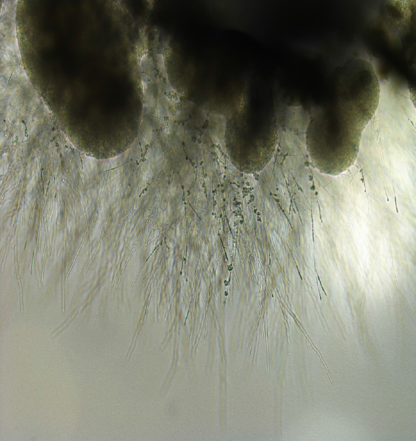
Figure 4 Appearance of Exophiala dermatitidis under stereoscopic microscope.
Sputum sample of pneumonia patient from our institution. Melanized, dimorphic, dematiaceous, and hyphal-growth-state fungus, with multiple conidial forms, was confirmed.
- Citation: Usuda D, Higashikawa T, Hotchi Y, Usami K, Shimozawa S, Tokunaga S, Osugi I, Katou R, Ito S, Yoshizawa T, Asako S, Mishima K, Kondo A, Mizuno K, Takami H, Komatsu T, Oba J, Nomura T, Sugita M. Exophiala dermatitidis. World J Clin Cases 2021; 9(27): 7963-7972
- URL: https://www.wjgnet.com/2307-8960/full/v9/i27/7963.htm
- DOI: https://dx.doi.org/10.12998/wjcc.v9.i27.7963


