Copyright
©The Author(s) 2021.
World J Clin Cases. Sep 16, 2021; 9(26): 7863-7869
Published online Sep 16, 2021. doi: 10.12998/wjcc.v9.i26.7863
Published online Sep 16, 2021. doi: 10.12998/wjcc.v9.i26.7863
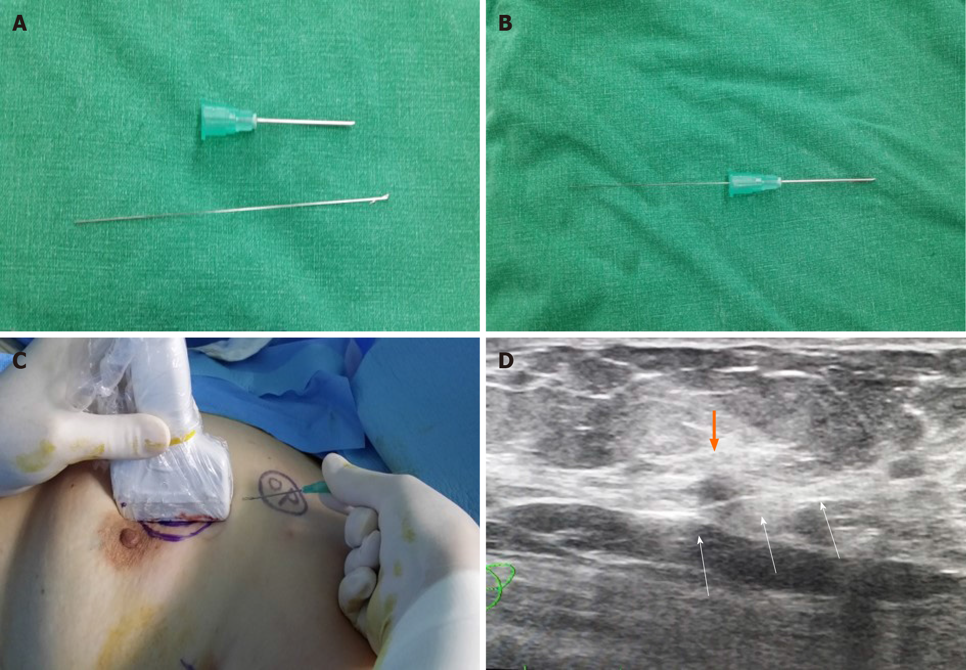
Figure 2 Method of intraoperative ultrasound-guided wire localization before operation.
A and B: The breast lesion localization wire consisted of a 23-gauze needle, through which a 25-gauze, 10 cm long monofilament wire with a distal hook; C and D: The wire was inserted and left in the breast as the needle was totally withdrawn. The three white arrows indicates the localized wire (white arrows) that was inserted to left breast mass (orange).
- Citation: Choi YJ. Migration of the localization wire to the back in patient with nonpalpable breast carcinoma: A case report. World J Clin Cases 2021; 9(26): 7863-7869
- URL: https://www.wjgnet.com/2307-8960/full/v9/i26/7863.htm
- DOI: https://dx.doi.org/10.12998/wjcc.v9.i26.7863


