Copyright
©The Author(s) 2021.
World J Clin Cases. Sep 16, 2021; 9(26): 7857-7862
Published online Sep 16, 2021. doi: 10.12998/wjcc.v9.i26.7857
Published online Sep 16, 2021. doi: 10.12998/wjcc.v9.i26.7857
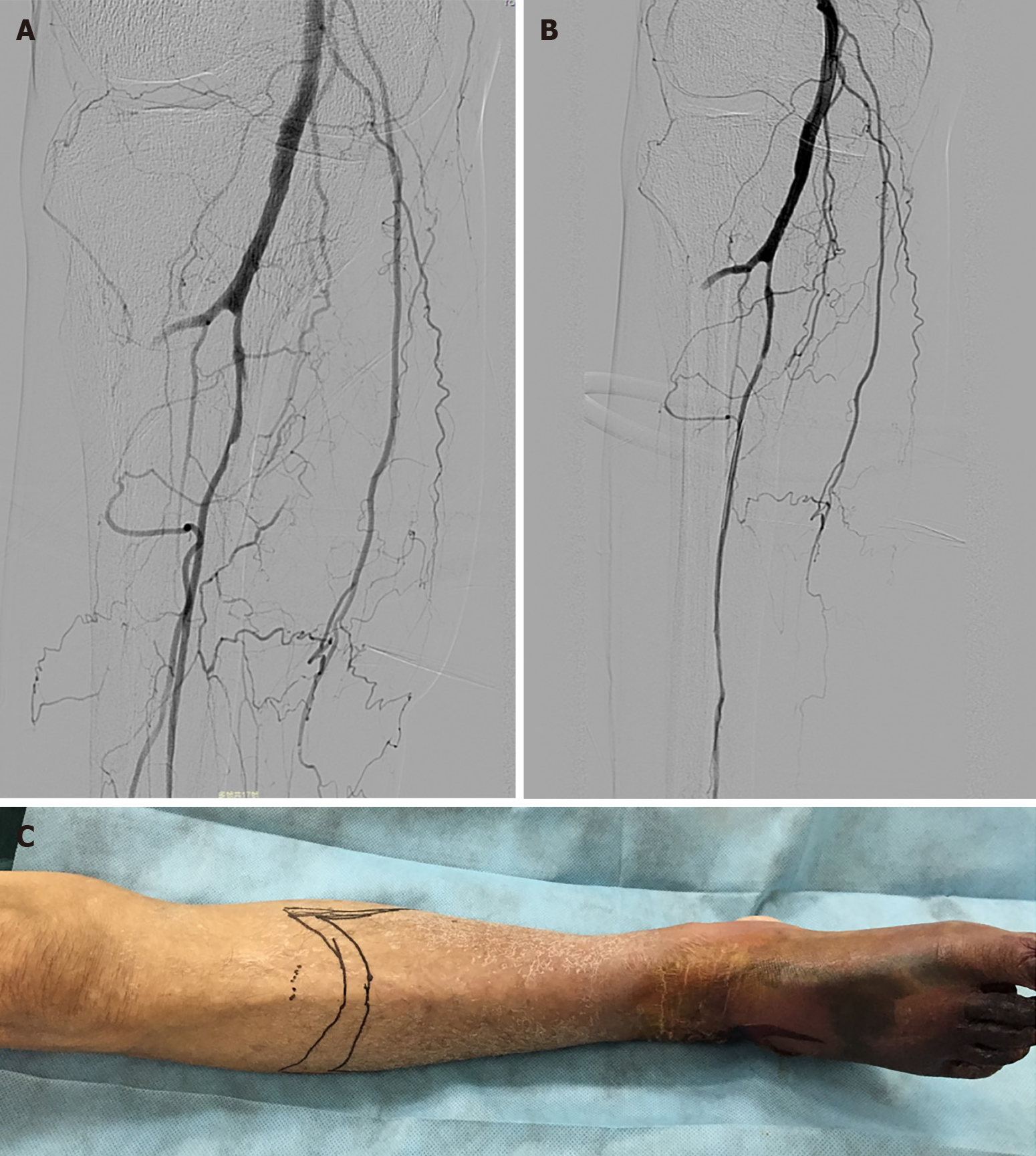
Figure 3 Angiography and physical examinations of the right anterior tibia.
A: On the sixth day of thrombolytic therapy, the proximal right anterior tibial and peroneal arteries were imaged by angiography; B: The right anterior tibial artery and peroneal artery were treated by low-pressure balloon dilatation; C: The amputation level was 9 cm below the right tibial plateau (marked with a marker pen).
- Citation: Lu ZY, Wang XD, Yan J, Ni XL, Hu SP. Critical lower extremity ischemia after snakebite: A case report. World J Clin Cases 2021; 9(26): 7857-7862
- URL: https://www.wjgnet.com/2307-8960/full/v9/i26/7857.htm
- DOI: https://dx.doi.org/10.12998/wjcc.v9.i26.7857


